
Section 1
Preview this deck
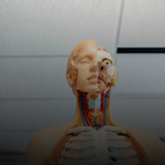
What is the basal nuclei?
Front

| 5 | 0 | ||
| 4 | 0 | ||
| 3 | 0 | ||
| 2 | 0 | ||
| 1 | 0 |
Active users
0
All-time users
0
Favorites
0
Last updated
6 years ago
Date created
Mar 1, 2020
Cards (142)
Section 1
(50 cards)
What is the basal nuclei?
Collection of masses of Grey matter in the white matter of the brain and contain neurons: caudate nucleus, lentiform nucleus, and amygdala Includes: 1) Corpus striatum 2) Amygdaloid nucleus 3) Claustrum FUNCTION: cooperate with the cerebral cortex in controlling movements - Receive input from many cortical areas Evidence shows that they: start, stop, and regulate intesnity of voluntary movements
Perineum
Inferior to the levator ani (pelvic diaphragm) that contains: - Males: penis, scrotum, and anus - Females: external genitalia (vulva) and anus
Sensory areas of the cerebral cortex
Cortical areas involved in conscious awareness of sensation that is located in the parietal, temporal, and occipital lobes - Distinct area for each of major senses located along the postcentral gyrus - Corresponds to the Brodmann areas 1-3 - Involved with conscious awareness of general somatic senses Projection is contralateral Sensory homunculus = body map of sensory cortex
The nerve impulse
- Generated at the initial segment of the axon and the conducted along it and releases neurotransmitters at the axon terminals - The neurotransmitters can excite or inhibit neurons - Neurons receive and send signals
Pelvic outlet
Between tip of coccyx and lower border of pubic symphysis
What are the five essential components of the reflex arc?
1) Receptor = site where stimulus acts 2) Sensory neuron = transmits afferent impulses to the CNS 3) Integration center = consists of one or more synapses in the CNS 4) Motor neuron = conducts efferent impulses from integration center to an effector 5) Effector = muscle or gland cells - Responds to efferent impulses - Contracting or secreting
What is the corpus striatum
Situated lateral to the thalamus and divided by a band of nerve fibers called the internal capsule into the caudate nucleus and lentiform nucleus
Lumbosacral plexus
Group of PNS spoinal nerves - Responsible for cutaneous and muscular innervation of lower limbs, parts of abodmen and pelvis - Derives from the ventral rami of T12-S4 Lumbar plexus = T12-L4 (three major nerves are genitofemoral, lateral femoral cutaneous, femoral nerves - innerv. abdominal, thigh, and hip musculature/skin) Sacral plexus = L4-S4 = sciatic and pudendal nerve from this - sciatic to femur and branches at knee , pudental nerve innervates the muscles of perineum and skin of genitalia
What are the muscular walls of the pelvis?
- Boney pelvis is separated inferiorly from perineum by levator ani muscle - Ala is covered by iliacus muscle accompanied by psoas major (iliopsoas) muscle Lateral wall: - Covered by obturator internus muscle arising from pelvic surface of ilium and ischium beneath the pelvic brim Posterior wall: - Piriformis Pelvic floor: - Levator ani and coccygeus (ischiococcygeus)
False vs true pelvis
Superior part = false - Anterior wall = muscular Inferior = True (Childbearing) - Anterior wall = bony Border between them is the SUPERIOR PELVIC APERTURE (pelvic brim or inlet) that is between the promontory of the sacrum and pubic tubercle
Sensory (afferent) signals
Picked up by sensor receptors and carried by nerve fibers of PNS to the CNS Includes somatic and visceral sensory
Cranial nerves list
I - Olfactory - S II - Optic - S III - Oculomotor - M IV - Trochlear - M V - Trigeminal - M VI - Abducent - M VII - Facial - S&M VIII - Vestibulocochlear - S IX - Glosssopharyngeal - S&M X - Vagus - S&M XI - Accessory - M XII - Hypoglossal - M
Visceral motor sensory
General: motor innervation of smooth muscle, cardiac muscle, and glands; equivalent to autonomic nervous system (ANS)
Lentiform nucleus
composed of the Globus pallidus and Putamen
What are the cells of the nervous system?
There are two main cell types: 1) Neurons - transmit electrical signals, found in the gray matter of the CNS and ganglia 2) Neuroglial cells (support cells) - nonexcitable, surround and wrap neurons
Somatic sensory
General: touch, pain, pressure, vibration, temperature, and proprioception in the skin, body wall and limb Special: hearing, equilibrium, vision, and smell
What are synapses
The site at which neurons communicate, signals pass along the synapse in one direction Presynaptic neuron = conducts signal toward a synapse Postsynaptic neuron = transmits electrical activity away from a synapse
What is the motor area of the cerebral cortex?
- Specific pyramidal cells that control specific areas of the body Face and hand muscles are controlled by many pyramidal cells Motor homunculus = body map of the 'motor cortex' Somatotopy = body is represented spatially in many parts of the CNS
Visceral sensory
General: stretch, pain, temperature, chemical changes, and irritation in viscera, nausea and hunger! Special: taste
Internal iliac artery
Main artery of pelvis Originates from common iliac - Passes downward to greater sciatic foramen - Eventually supplies gluteal and pelvic muscles
Somatic motor sensory
General: motor innervation of all skeletal muscles (except the pharyngeal arch muscles)
motor (efferent) signals
carried away from the CNS and innervate muscles and glands Includes somatic motor, branchial motor, and visceral motor
Branchial motor sensory
Motor innervation of the pharyngeal arch muscles
What are the four regions of the brain?
1) Cerebral hemispheres 2) Diencephalon 3) Brain stem: midbrain, pons, and medulla 4) Cerebellum
The pelvis is separated from the perineum by the
Pelvic diaphragm, which is the levator ani muscle
What are the cerebral meninges?
- Both brain and spinal cord are covered - Dura, arachnoid, and pia maters
What are neurons?
The basic structural units of the nervous system and possess a cell body and processes called neurites - Human body consists of billions
CSF (Cerebrospinal fluid)
The brain and spinal cord are suspended in it CSF is in the subarachnoid space
What is the primary motor cortex?
Controls motor functions - Primary motor cortex = somatic motor area - Located in the precentral gyrus (Broadmann area 4) - Pyramidal cells = large neurons of primary motor cortex Corticospinal tracts descend through the brainstem and spinal cord - Axons signal motor neurons to control skilled movements Contralateral = the pyramidal axons cross over to the opposite side of the brain
What are the ventricles in the brain?
Expansions of the brain's central cavity that are filled with CSF, lined with ependymal cells and continuous with each other - Continuous with the central canal of the spinal cord 1) Lateral ventricles - Located in cerebral hemispheres, horseshoe shape 2) Third ventricle - lies in diencephalon and connected with lateral ventricles by interventricular formen of Monro 3) Fourth ventricle - lies in the hindbrain, connects to the central canal of the spinal cord 4) Cerebral aqueduct of Sylvius = connects 3 and 4th ventricle
Cranial nerves
12, leave the brain and pass through foraminae and fissures in the skull All are distributed in the head and neck, except for the 10th that also supplies the thoracic and abdominal structures
Brodmann areas
What is the cerebellum?
Located dorsal to the pons and medulla - Smoothes and coordinates body movements and maintain equilibrium - Has two cerebrellar hemispheres that are each divided into: anterior, posterior, and flocculonodular lobe - Surfaces fold into ridges called folia that are separated by fissures
What are the types of neuroglial cells in the CNS?
1) Astrocytes - most abundant glial cell type that take up and release ions to control the environment around neurons, recapture adn recycle neurotransmitters 2) Microglia - smallest and least abundant - phagocytes, the "macrophages of the CNS" 3) Ependymal cells - line the central cavity of the spinal cord & brain 4) Oligodendrocytes - produce myelin sheaths in CNS
Excitatory vs inhibitory synapses?
Excitatory: - Depolarizes the postsynaptic membrane Inhibitory: - Reduces the ability of the postsynpatic neuron to generate an action potential
Divisions of the nervous system?
Central & Peripheral nervous system
What are the cerebral hemispheres components
Fissures = deep grooves that separate the major regions of the brain - Transverse fissure = separates cerebrum and cerebellum - Longitudinal fissure = separates cerebral hemispheres Sulcus = grooves on the surface of the cerebral hemispheres - Deeper suli divide cerebrum into lobes (which are named for the skullbones overlying them) Gyrus = twisted ridges between the sulci
What is the spinal cord?
- Covered by meninges and is suspended in the CSF Segments (structure): 1) Cervical (C) = 8 2) Thoracic (T) = 12 3) Lumbar (L) = 5 4) Sacral = 5 5) Coccygeal = 1 Conus medullaris Cauda equina Filum terminale
What are the sulci of the cerebral hemispheres?
Central sulcus = separates frontal and parietal lobes, and is bordered by two gyri the precentral gyrus and postcentral gyrus Parieto-occipital sulcus = separates the occipital from the parietal lobe Lateral sulcus = separates temporal lobe from parietal frontal lobes Insula = deep within the lateral sulcus
What is the somatosensory assocaition area (sensory of cerebral cortex?)
Lies posterior to the primary somatosensory cortex - Corresponds to the Brodmann areas 5 and 7 - Integrates different sensory inputs: touch, pressure, and others Draws upon stored memories of past sensory experiences
What are the reflex arcs?
A simple chain of neurons - Determine the structural plan of the nervous system - Responsible for reflexes - They are rapid, autonomic nervous responses and can be either visceral or somatic
What are the three overlapping functions of the nervous system?
1) Receives inputs from outside and inside the body 2) Processes and interprets sensory input - makes decisions/integration 3) Dictates a response by activating effector organs - response: motor output, muscle contraction, or glandular activity
What are the connective tissues covering the nerves?
1) Endoneurium = layer of delicate connective tissue surrounding the axon 2) Nerve fascicles - groups of axons bound into bundles 3) Perineurium - connective tissue wrapping surrounding a nerve fascicle 4) Epineurium - whole nerve is surrounded by tough fibrous sheath
What is the cerebral cortex?
- "conscious mind" that enables self-awarness and our sensations - They initiate and control voluntary movements, commuincate, remember, and understand - Composed of gray matter neuronal cell bodies, dendrites, and short axons - Folds in cortex, triples its size - Has Brodmann areas that are 52 structurally distinct areas
What is the action potential?
- A strong stimulus is applied to the axon triggers nerve impulse or action potetnail - The membrane becomes negative externally and the impulse travels the length of the axon - The membrane repolarizes itself!
What is the brain stem?
Includes the midbrain, pons, and medulla oblongata - General functions: produces autonomic behaviors necessary for survival - Passageway for all fiber tracts running BETWEEN CEREBRUM and spinal cord - Heavily involved with innervation of face/head - 10 of the 12 pairs of cranial nerves are attached to it (I and II don't)
What are the types of neuroglial cells in PNS?
1) Satellite cells - surround neuron cell bodies within ganglia 2) Schwann cells - surround axons in the PNS, form myelin sheath around axons of the PNS
What are the functional regions of the cerebral cortex?
- Regions of cerebral cortex that perform distinct motor and sensory functions, the memory and language spread over wide area Three kinds: 1) Motor areas 2) Sensory areas 3) Association areas
What are the types of reflexes?
monosynaptic: - Simplest - Just one synapse, and the fastest of all - EX: knee-jerk Polysynaptic - More common - Most have a single interneuron between the sensory and motor neuron - EX: withdrawal reflexes
What are the neuroglia (supporting cells)?
There are 4 in the CNS, and 2 in the PNS - They provide supportive functions for neurons - They cover the nonsynaptic regions of neurons
Section 2
(50 cards)
The mucosa of the uterus
"Endometrium" - Lies directly on the muscle (myometrium) - Has simple high columnar epithelial (some ciliated) cells and contain uterine glands - There is a basal layer (not shed during menstruation) that can be distinguished from a functional layer stratum compactum and St. spongiosum up to 8mm
What happens to the uterine muscles during pregnancy?
The undergo hyperplasia/hypertrophy and then regress
Blood supply of the testis?
Testicular (gonadal) arteries from the abdominal aorta VEINS: - Blood from epididymis and testis gets into pampiniform plexus and from there to the testicular vein - The left testicular vein drains into the left renal vein; and the right testicular vein drains directly into the IVC
What are the layers of the scrotum?
1) Skin 2) Dartos fascia - contains dartos muscle that is attached to skin of scrotum and thereby its contraction causes scrotum to wrinkle when cold to prevent heat loss 3) External spermatic fascia 4) Cremaster muscle and fascia 5) Internal spermatic fascia 6) Tunica vaginalis
What is the broad ligament of the uterus?
Mesometrium, ligamentum latum uteri - The sheath of the peritoneum that is lifted by the uterus and Fallopian tubes Mesosalpinx and mesovarium are folds of the broad ligament - The Vesicouterine and rectouterine (douglas) pouches are forward/backward extensions of the broad ligament from the uterus, over the urinary bladder, and rectum anterior and posterior to the uterus
A retroverted uterine may be a cause of
Abortion/dyspareunia
The internal Os of the cervix, Cervical canal, and external Os is ___ in Nulliparous and ___ after childbirth
Round; transverse
What are the ovaries?
- 2.5-5cm long and 0.5-1cm thick - Covered by mesovarium - attached to the uterus by means of the ligament of the ovary (ovarian ligament), which arises from the posterior surface of the lateral angle of the uterus - Ovary is attached to the lateral abdominal (and pelvis) wall by means of the suspensory ligament of ovary, which contains the ovarian artery and vein
Pap smear
Counts various cell types: PBC SSC ISC
What is the secretion phase?
Day 15-28 - Controlled by progesterone; mucous secretion and increase in blood vessels - At the end of the phase, progestrone decreases and arteries contract due to drying - Ischemia follows and tissue damage and bleeding starts again
Ectopic pregnancy
If implantation occurs at places other than the anterior or posterior walls of the body of uterus - The uterine tube cannot accomodate the place for pregnancy beyond the 2nd month - Causes severe pain (acute abdomen), surgical evacuation
Lymphatics of scrotum
To the superficial inguinal lymph nodes
Histerosalpingography
Radioopaque material is injected into the uterus to check for the shape and anomalies of the uterus and patent tubes
Innervation of the testis
Sympathetic from T7 Parasympathetic from Vagus coming along the testicular artery as testicular plexus
What are the internal genital organs (female)?
- Ovaries - Uterine (Fallopian) tubes - Uterus (womb) - Vagina situated within the lesser pelvis
What are the fallopian tubes?
- Run from the tubal angle of the uterus to the surface of the ovary (on abdominal end) - Intraperitoneal and mobile - fixed by the mesosalpinx
Innervation to male urethra
- pudendal nerve, pelvic splanchnic nerves, and sympathetic system
What is the uterus?
- Anchored between urinary bladder and rectum by muscular connective tissue retinaculum in the subperitoneal connective tissue - Also fixed by the round ligament, which arises from the anterior aspect of the lateral angle (Ovarian ligament comes from the posterior aspect) - and passes through the inguinal canal to reach labia major
Lymphatic drainage of testis
To the preaortic and lumbar lymph nodes
Loss of support of uterus may cuase
(Due to several birthds, old age), - Uterus may drop lower or its portio part comes out of the vagina (prolapse)
WHat is the pampiniform venous plexus?
A thermoregulator of the testis to keep the temperature constant
Blood supply of the Fallopian tube
By the ovarian and uterine artery
What is the blood supply of the scrotum?
1) Internal pudendal artery to the posterior scrotal artery 2) External pudendal artery after giving anterior scrotal artery 3) Cremasteric branch of inferior epigastric artery VEINS SAME
Where do ejaculatory ducts open?
On or near the prostatic utricle
What is the spongy urethra?
15-16cm long - Starts from the bulb of the penis and ends at the external urethral orifice of the glans penis
How is the uterus supported?
Dynamic and passive support 1) Dynamic support: - given by *pelvic diaphragm (the levator ani and coccygeus muscle) 2) Passive (mechanical support) - Uterus is normally anteflexed and bent forward over bladder 90 degrees - Responsible for passive support and prevents it from being pushed out of genital opening
What is the male urethra?
Muscular tube about 20 cm long - starting from the internal urethral orifice of the urinary bladder to the external urethral orifice at the tip of the glans penis Has four parts: 1) Preprostatic 2) Prostatic 3) Membranous 4) Spongy parts
Blood supply of the uterus
Uterine artery (from internal iliac) Ovarian artery Some from vaginal artery
Blood suppy to female urethra
Internal pudendal and vaginal arteries and (Internal pudendal and vaginal veins)
Testis
"Orchis" - Suspended in scrotum by spermatic cord - Left testis hangs lower - Tightly enveloped by Tunica albuginea capsule - Septate divide the testicle into 200-300 lobules - Each lobule contains many seminiferous tubules that lead into the Rete testis with straight endings: Tubuli recti - The tubuli recti are interconnected and lead to efferent ductules - then reach the ducts (lobules) of epididymis that will merge into ductus deferens This is the pathway - spermatozoa produced in the lumen of the seminiferous tubule are transported through this pathway to the prostate and urethra
What is in the prostatic part of the male urethra?
4cm long is inside the prostate - The most dilated part of the urethra - Ends where the urethra is covered by the external urethral sphincter - Has the urethral crest on the posterior wall on both sides of which are the prostatic sinuses into which most prostatic ductules open
Uterus
- Divided into body, neck, and fundus - In a Nullipara adult lady it is 7.5 long, 5 cm wide, and 2.5 thick - It is composed of smooth muscle = myometrium, mucosa - Endometrium (mucosa) - simple high columnar epithelium Fundus = part of uterus rising above the tubal angle Neck = Cervix (2.5cm) - Includes supravaginal and vaginal part ("Portio") - Portio is covered by vaginal epithelium that is stratified squamous non-keratinizing epithelium Isthmus (lower segment)
Female urethra
4-6 cm long starting from the internal urethral orifice to the external urethral orifice in the vestibule of vagina, anterior to the vaginal opening - Urethra accompanied by the vagina pass through the pelvic diaphragm and perineal membrane - Surrounded by the external urethral sphincter - INferior part of urethra is in the perineum - Urethral glands open into urethra (mostly upper) - Paraurethral glands = homologous to prostate
What is the Fallopian tube?
- 8-20 cm long - Lie intraperitoneally in the mesosalpinx of the broad ligament Has four parts: 1) Infundibulum 2) Ampulla 3) Isthmus 4) Uterine part
Urinary bladder
Lies in the lesser pelvis in adults, beneath the peritoneum behind the pubic bone (newborn upper than the pubic bone) - The median umbilical ligament is remnant of Urachus and ascends from its vertex - Medial umbilical ligament (obliterated umbilical arteries) are found around its body extended to the umbilicus - Both ureters enter the bladder at the base of the Trigone and the urethra leaves it from there - Has transitional type epithelium and the mucosa is soft-reddish in life
What is the ligamentous support of uterus?
Cervix of the uterus is the least mobile part of uterus because it is supported by endopelvic fascia or Retinacula (that also contains smooth muscle) 1) Transverse cervical (cardinal) ligament = extended from cervix to lateral pelvic walls 2) Sacrocervical (sacrouterine) lig - posteriorly 3) Pubocervical (pubouterine) lig anteriorly 4) Round ligament = gives suport to uterus too
Menstrual cycle
Commonly 28 days (counting first day of bleeding) 1) Phase of desquamation and regeneration (A) = 1st - 4th day 2) Phase of proliferation (B,C) = 5th -15th day 3) Phase of secretion (D,E) = 15h-28th day
Incompetent internal Os
Leads to habitual abortion before the 3rd month Requires tying of cervix = Shirodka/McDonald
What are the four parts of the Fallopioan tube
1) Infundibulum - open with finger-like fimbriae to catch egg 2) Ampulla - dilated part of tube (5-6 cm) which has longitudinal grooves to guide ovum 3) Uterine part - opens into the lumen of uterus 4) Mucosa - Branching folds and is simple high columnar ciliated epithelium (pseudostratified) with glandular cells - Some fluid is produced here and the celiae produce a current toward the uterus to help migration/distribution of spermatozoa, muscular layer (peristalsis toward uterus) After the ovum is released from the follicle, it reaches the funnel via fimbriae with 3-6 mins
What is the desquamation/regeneration phase of menstruation?
Day 1 -4 Disappearance of progesterone and increase in estrogen - Superficial portion of Endometrium is shed and eventually the epithelium and connective tissue of the functional layer regenerate from the basal layer and wound is closed
Innervation of the uterus
Sympathetic and parasympathetic Pelvic splanchnic nerves - S2-S4
Urinary bladder volumes
Can hold over 700 mL of urine voluntarily - however the urge is around 350 mL unless we don't do it voluntarily
Blood supply of urethra
Inferior vesicle and middle rectal arteries and the internal pudendal artery to the lower parts SAME NAMES FOR VEINS
What is the site of fertilization in the Fallopian tube?
The ampulla, NOT the site of implantation Implantation occurs in the uterus -- the cilia pass the fertilized egg toward the uterus
Salpingitis
An inflammation (Pelvic Inflammatory Disease) and infection (gonorrhea and chlamydia) of the Fallopian tubes that may lead to loss of the epithelium - which may interfere with fertilization leading to sterility
What are Leiomyomas (Fibroids)?
- Most common overall tumors in women - In uterus - More common in blacks than whites - Estrogen-sensitive smooth muscle tumors that in pregnancy may enlarge and cause obstructive delivery
Innervation of the scrotum
1) Genitofemoral = anterolateral 2) Ilioinguinal (anterior) 3) Pudendal (posterior) 4) Posterior femoral cutaneous nerve (inferior)
What is the proliferation phase of menstruation?
Day 5- 15 - Ovulation period - Mainly controlled by estrogen - The functional layer grows, glands get bigger, spiral arteries form, and body temperature rises OVULATION USUALLY TAKES PLACE 13-14 DAYS BEFORE FIRST DAY OF NEW MENSTRUAL PERIOD
What is the membranous (intermediate) part of the male urethra?
- Part surrounded by the external urethral sphincter and perineal membrane - Posterolateral to this part of the urethra are the Cowper's (Bulbourethral glands) that open into the spongy urethra - The most vulnerable part to injuries due to not being fixed/less mobile
What are the external genital organs (female)?
Labia major and labia minor and clitoris Vestibule of the vagina and vestibular glands
Section 3
(42 cards)