
Section 1
Preview this deck
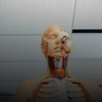
aortic arch
Front

| 5 | 0 | ||
| 4 | 0 | ||
| 3 | 0 | ||
| 2 | 0 | ||
| 1 | 0 |
Active users
0
All-time users
0
Favorites
0
Last updated
6 years ago
Date created
Mar 1, 2020
Cards (127)
Section 1
(50 cards)
aortic arch
coronary arteries and veins
smooth muscle
internal carotid
branches off the common carotid and runs inward and upward to supply the brain
valves
only present in veins and lymph nodes, prevents back flow
Pericardium
skeletal muscle
thoracic aorta
part of the aorta the descends from the aortic arch through the thorax to the diaphragm
Myocardium
muscular, middle layer of the heart
Endocardium
inner lining of the heart
ventricles
Lower chambers of the heart
external elastic membrane
separates tunica media from tunica externa
external carotid artery
Artery that supplies blood to the anterior (front) parts of the scalp, ear, face, neck, and sides of the head.
right common carotid artery
descending aorta
papillary muscles
responsible for pulling the atrioventricular valves closed by means of the chordae tendineae
superior vena cava
sheep ventricles
internal elastic membrane
separates the tunica intima from the tunica media
tunica externa
interatrial septum
separates atria
left common carotid artery
semilunar pulmonary valve
The heart valve between the right ventricle and the pulmonary artery that controls blood flow from the heart into the lungs.
Pericardium
Membrane surrounding the heart
semilunar aortic valve
between left ventricle and aorta
epicardium
outer layer of the heart
pulmonary trunk
tunica intima
includes a thin layer of endothelium and a basement membrane
interventricular septum
separates ventricles
sheep atria
intercalated disks
strong junctions between cells and immediate transmission from one cell to another
ascending aorta
Base of heart
tricuspid valve
valve between the right atrium and the right ventricle
vasa vasorum
small vessels that supply blood to outer part of the larger vessels (tunica externa)
sheep auricles
bicuspid valve
valve between the left atrium and the left ventricle.
trabeculae carneae
muscular ridges on the internal surface of the ventricles
systemic circuit of heart
left side of the heart, which pushes oxygen rich blood through the aorta of the heart to the tissues and back to the heart
pulmonary arteries
epicardium
D
tunica media
pulmonary trunk
pulmonary circuit
right side of the heart which receives carbon dioxide rich blood form tissues and send the blood to the lungs
Atria
upper chambers of the heart
pulmonary veins
chordae tendineae
thin bands of fibrous tissue that attach to the valves in the heart and prevent them from inverting
inferior vena cava
cardiac muscle tissue
apex
Section 2
(50 cards)
axillary atery
facial artery
intercostal arteries
ulnar artery
superior vena cava
celiac trunk
renal artery
left subclavian artery
popliteal artery
common iliac vein
celiac trunk branches
gastric artery, splenic artery, common hepatic artery
superior mesenteric vein
drains small intestines and ascending colon
internal iliac vein
vertebral artery
cephalic vein
Adominal Aorta
gonadal artery
external iliac artery
brachial artery
femoral vein
superior mesenteric artery
large abdominal artery; feed large and small intestine
right subclavian artery
ulnar vein
hepatic vein
drains the liver
lumbar vein
a group of veins that drain the posterior abdominal wall, vertebral column and spinal cord and return to the inferior vena cava
intercostal vein
external jugular vein
inferior mesenteric artery
supplies large intestine
vertebral vein
E
lumbar artery
four paired branches off the posterior side of the descending aorta, supply lumbar muscles and spinal cord
brachial vein
external iliac vein
radial vein
basilic vein
brachiocephalic vein
femoral artery
internal iliac artery
subclavian vein
facial vein
drains facial structures
brachiocephalic trunk
renal vein
gastric vein
common iliac artery
splenic vein
radial artery
internal jugular vein
gonadal vein
veins of hepatic portal system
splenic, gastric, superior mesenteric, inferior mesenteric, hepatic portal, hepatic
inferior mesenteric vein
drains distal portion of large intestine
hepatic portal vein
carry blood full of nutrients and other substances from the gastrointestinal tract and spleen to the liver
Section 3
(27 cards)