AP II Heart Anatomy Pt. 1

AP II Heart Anatomy Pt. 1

memorize.aimemorize.ai (lvl 286)
Section 1

Preview this deck

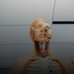
Where's the anatomical location of the heart?

Front
Star 0%
Star 0%
Star 0%
Star 0%
Star 0%

0.0

0 reviews

5
0
4
0
3
0
2
0
1
0

Active users

0

All-time users

0

Favorites

0

Last updated

6 years ago

Date created

Mar 1, 2020

Cards (42)

Section 1

(42 cards)

Where's the anatomical location of the heart?

Front
the transport system pump, enclosed within the mediastinum

Back

tricuspid valve

Front

Back

What is a serous membrane?

Front

A thin, continuous membrane lining a closed cavity of the body and covering its organs -parietal and visceral layers

Back

anterior intraventricular artery

Front

the red one

Back

Similarities between cardiac and skeletal muscle

Front

-striated -role of calcium & ATP

Back

trabeculae carneae

Front

Back

right pulmonary artery of the heart

Front

Back

aortic arch

Front

Back

left pulmonary veins

Front

Back

Differences between cardiac and skeletal muscle

Front

cardiac: -short, flat, branched, interconnected, 1 or 2 nuclei per cell -gap junctions -gap junctions create functional syncytium -large mitochondria -sarcomeres contain less extensive banding pattern skeletal: -striated, long, cylindrical, multinucleate -no gap junctions between cells -fewer mitochondria

Back

left ventricle of the heart

Front

Back

apex of the heart

Front

Back

right coronary artery

Front

Back

What is the function of syncytium?

Front

cardiac muscle cells are electrically connected by gap junctions and thus the entire myocardium behaves as a single unit

Back

left atria

Front

Back

what is the function of intercalated discs?

Front

1. help to maintain the three-dimensional structure of the heart 2. forms a bridge linking cells together so they all pull as a unit 3. allows for direct electrical connections of all cells location where cardiac plasma membranes interlock

Back

What are the three layers of the heart wall?

Front

epicardium (visceral pericardium): external layer myocardium: middle layer endocardium: inner layer

Back

bicuspid valve

Front

Back

right pulmonary veins

Front

stick out of superior vena cava

Back

What is the pathway of blood through the heart?

Front

1. superior vena cava, inferior vena cava, coronary sinus 2. right atrium 3. tricuspid valve 4. right ventricle 5. pulmonary semilunar valve 6. pulmonary trunk to lungs to heart 7. 4 pulmonary veins 8. left atrium 9. mitral valve 10. left ventricle 11. aortic semilunar valve 12. aorta

Back

posterior intraventricular artery

Front

the blue one

Back

superior vena cava

Front

Back

what is the sac that encloses the heart?

Front

pericardium: double-walled -deep to fibrous pericardium= serous pericardium (parietal and visceral layers)

Back

left coronary artery

Front

Back

auricles of the heart

Front

Back

What are the AV valves?

Front

tricuspid (right) and bicuspid (left)

Back

What is collateral circulation?

Front

The development of new, smaller branches of coronary arteries that grow in response to a vessel that has slowly become occluded or nearly occluded over time.

Back

ascending aorta

Front

Back

inferior vena cava

Front

Back

papillary muscle of the heart

Front

Back

chordae tendineae

Front

Back

What is PMI?

Front

point of maximal impulse

Back

fossa ovalis

Front

Back

intraventricular septum

Front

Back

right atria

Front

Back

right ventricle of the heart

Front

Back

coronary sinus

Front

Back

what is the SL valve operation?

Front

opening of SL valves: as ventricles contract and intraventricular pressure rises, blood is pushed up against SL valves, forcing them open closing of SL valves: as ventricles relax and intraventricular pressure falls, blood flows back from arteries, filling the cusps of SL valves and forcing them to close

Back

What is the AV valve operation?

Front

opening of AV valves: 1. blood returning the the heart fills the atria, pressing against the AV valves. the increased pressure forces AV valves open 2. as ventricles fill, AV flaps hang limply into ventricles 3. atria contract, forcing additional blood into ventricles closing of AV valves: 1. ventricles contract, forcing blood against AV valve cusps 2. AV valves close 3. papillary muscles contract and chordae tendinae tighten, preventing valve flaps from everting into atria

Back

pectinate muscles

Front

Back

pulmonary trunk

Front

Back

left pulmonary arteries

Front

Back