
Section 1
Preview this deck
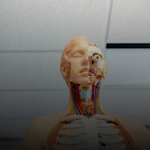
Eccentric Contraction
Front

| 5 | 0 | ||
| 4 | 0 | ||
| 3 | 0 | ||
| 2 | 0 | ||
| 1 | 0 |
Active users
2
All-time users
2
Favorites
0
Last updated
1 year ago
Date created
Mar 1, 2020
Cards (603)
Section 1
(50 cards)
Eccentric Contraction
-Muscle contracts as it is stretched -Counterintuitive -Generates up to 50% more force than a concentric contraction -Tissue damage possible
Maximal Stimulus
-Muscle contractions increase in force with increasing stimulus until they reach this
Myofibrils
-Skeletal muscle -a rod like structure -full of sarcomeres -contractile unit of muscle
Actin
-Thin filaments, anchored to the Z disc -These "heads" serve as cross bridges -Binding sites: ATP Actin ATPase enzyme
Hyperplasia
-Adding more muscle fibers, not common
Isometric Contraction
-Tension develops, but no shortening -Load>tension
Sliding Filament Contraction Model
-Thin filaments are culled by thick filaments -Z discs pulled towards the center -I bands shorten -H zones disappear
Muscle Tension
-The force exerted by a contracting muscle
Load
-Force exerted on a muscle
Muscle Function
-Movement of bones, digestive tract, blood vessels -posture and body position -stabilize joints -heat generation
Myofilaments
-Actin -Myosin
Insertion
-Movable location
Direct Attachment
-Epimysium of muscle fuses to the periosteum or the bone or the perichondrium of the cartilage
Unipennate
-Fascicles insert from opposite sides
Sarcolemma
-Skeletal muscle -Cell membrane
Types of Muscle Tissue
-Skeletal -Smooth -Cardiac
Indirect Attachment
-Sheaths extend beyond muscle -Attach to periosteum via tendon or aponeurosis
Muscle Arrangement Circular
-Fascicles arranged in concentric rings -Orbicularis oris
Cardiac Muscle
-Only in heart -Striated and branched -Involuntary contractions, does not need nervous system
Concentric Contraction
-Muscle shortens
Sliding Filament Relaxed Model of Contraction
-Filaments only overlap at edges of A band
Isotonic Contraction
-Tension>load -Two different types of length change
Myosin
-Thick filaments, connected at M line -Rod-like tail with two heavy myosin chains, note the "heads" on the myosin chains -These "heads"serve as cross bridges -Binding sites: ATP Actin ATPase enzyme
Sarcoplasm
-Skeletal muscle -Cytoplasm, glycogen storage and myoglobin for oxygen storage
Motor Units
-Made up of the motor neuron and the muscle fibers it supples; can be few or hundreds -Number of fibers per motor units relates to amount of control necessary (Fingers, eyes= small motor units. Hips, thighs= large motor units).
Muscle Arrangement Pennate
-Central tendon, short fascicles run at an angle to the tendon -Unipennate, Bipennate, Multipennate
Skeletal Muscle Fiber Physiology; Excitation-Contraction Coupling
-Generation and propagation of action potential along the sarcolemma -Triggered by rise in intracellular calcium
Muscle Arrangement Convergent
-Board origin, fascicles converge toward a single tendon insertion -Pectoralis major
Sarcomeres
-Region between the two Z discs -A contractile unto of muscle fibers
Muscles
-Movers of the body, uses nervous system and blood flow
Striations
-Myofibrils -Aligned series of A (dark) and I (light) bands -H zone, M line, Z disc
Smooth Muscle
-Walls of hollow organs (stomach, digestive tract, blood vessels, airways, uterus) -Not striated -Involuntary
Skeletal Muscle Fiber Physiology; Activation
-Neural stimulation at neuromuscular junction -From a signal called an action potential
Skeletal Muscle
-A single cells is a muscle fiber -Striated -Voluntary control, with rapid contractive and high force output -Nervous system stimulates -Lots of nuclei and mitochondria
Elastic Filaments
-Titin; holds thick filaments in place -Dystrophin; holds thin filaments to sarcolemma
Sarcoplasmic Reticulum
-Skeletal muscle -Endoplasmic reticulum of muscle -Smooth ER around each myofibril -Stores and regulates calcium -Terminal cisterns are perpendicular at A band/I band junction
Threshold Stimulus
-The strength at which the first observable muscle contraction occurs
Skeletal Muscle Fiber Physiology
-Contraction is regulated; Activation or Excitation-Contraction Coupling
Origin
-Non moving location
Characteristics of Muscles
-Excitability; respond to stimuli -Contractility; shorten forcibly when stimulated -Extensibility; ability to be stretched -Elasticity; ability to recoil to resting length
Hypertrophy
-Increase in the size of individual fibers (not number of muscle fibers), weight training (getting bigger) -More myofibrils and myofilaments
Muscle Arrangement Parallel
-Fascicles parallel to long-axis of muscle -Sartorius
Aponeurosis
-A sheet of connective tissue
Muscle Attachments
-Muscles attach in at last two location 1. Insertion 2. Origin
Sarcomeres in Parallel vs in Series
-Fibers in parallel generate a lot of force -Don't contract as far vs -Long fibers in series do a lot of shortening -Not a lot of force
Striations; Myofilaments Thick filaments Thin filament
-Create the banding pattern -Run the length of the A band -Run length of I band and into the A band
Muscle Arrangement Fusiform
-Spindle shaped muscles with parallel fibers -Biceps brachii
Sliding Filament Model of Contraction
-Generation of Force -Does not always mean fiber shortening -Shortening occurs when muscle tension exceeds the load opposing shortening
Botox
-Blocks ACH release
T-Tubules
-Inward extensions of sarcolemma at A band/I band junction -Triads formed by T-tubules and terminal cisternae
Section 2
(50 cards)
ATP
-The energy source for contraction -Depletes quickly -Replenishes three ways
Pupil
-Central opening that regulates amount of light entering eye -Close vision and bright light; sphincter pupillae -Distant vision and dim light; dilator pupillae -Changes in emotional state; pupils dilate when subject matter is appealing or requires problem-solving skills
Fatigue
-Physiological inability to contract muscle -Several different causes; >Ionic imbalance, messing with ECC >Shortage of metabolic reagents (glucose, glycogen, oxygen) all result in slowing ATP production -Not a lack in ATP
Cornea
-Bends light as it enters eye -Sodium pumps help maintain clarity of cornea -Pain receptors contribute to reflexes and blinking
Choroid Region
-Posterior portion of uvea -Supplies blood to all layers of eyeball -Brown pigment absorbs light to prevent light scattering and visual confusion
Lacrimal Apparatus
-Lacrimal gland and ducts that drain into nasal cavity -Lacrimal gland in orbit above lateral end of eye -Lacrimal secretion (tears)
Vison
-Has 70% of body's sensory receptors in the eye -Visual processing by 50% of the cerebral cortex -Protected by orbital bone and cushion of fat
Vascular Layer (Uvea)
-Middle pigmented layer -Three regions: choroid, ciliary body, and iris
Sclera
-Opaque posterior region -Protects, shapes eyeball; anchors extrinsic eye muscles -Continuous with dura mater of brain posteriorly
Lubricating Glands
-Tarsal Glands; Modified sebaceous glands that lubricate lid and eye -Ciliary Glands; Modified sweat glands, found between hair follicles
Six straplike extrinsic eye muscles
-Originate from bony orbit; insert on eyeball -Enable eye to follow moving objects; maintain shape of eyeball; hold in orbit
Third Class Lever body
-In most of the body -Effort is in the middle -LEF; tweezers, biceps
Sensory Organs
-Composed of localized, distinct, unique receptors in the head; Vision Taste Smell Hearing Equilibrium
Lever
-Levers result in a mechanical advantage -Ratio of each side of the lever around the fulcrum -Can be geared for speed or power
Two oblique muscles move eye
-Superior and inferior oblique muscles
Aerobic Pathway
-Produces 95% of ATP during rest and moderate exercise -Requires oxygen -Occurs in mitochondria -Generates glycogen, bloodborne glucose, pyruvic acid from glycolysis, and free fatty acids
Retina; Inner Neural Layer
-Transparent -Composed of three main types of neurons; Photoreceptors, bipolar cells, ganglion cells -Signals spread from photoreceptors to bipolar cells to ganglion cells -Ganglion cell axons exit eye as optic nerve
Eyelids
-Protect eye anteriorly -Meet at medial and lateral commissures
Asthma
-A smooth muscle disease
Humors Fluid
-Internal cavity filled with fluids
Eyelashes
-Nerve ending initiate reflex blinking
First Class Lever
-Fulcrum between the load and effort -LFE; seesaw, raising head
Muscle Metabolism
-Maintain force output, energy must be constantly supplied to the muscle
Anaerobic
-No oxygen -Produces less ATP -Glycolysis which produces lactic acid
Retina; Outer Pigmented Layer
-Single-cell-thick lining -Absorbs light and prevents its scattering -Phagocytize photoreceptor cell fragments -Stores vitamin A
Inner Layer; Retina
-Originates as out pocketing of brain -Delicate two layered membrane -Outer pigmented layer -Inner Neural layer
Accessory Structures fro Vision
Eyebrows Eyelids (palpebrae) Conjunctiva Lacrimal apparatus Extrinsic eye muscles
Lens separates internal cavity into
anterior and posterior segments (cavities)
Multipennate
-Appear as 'feathers' inserting into one tendon
Glycolysis
-Part of anaerobic respiration -Glucose degraded to 2 pyruvic acid molecules -Normally head to mitochondria -Changes at 70% of maximum contractile activity -Muscles squeezing out the blood -Pyruvic acid converted to lactic acid -Yields only 5% as much ATP as aerobic respiration, but 2.5 times faster
Second Class Lever
-Load is in the middle -FLE; wheelbarrel, toe raises
Tarsal plates
-Supporting connective tissue
Ciliary Body
-Smooth muscle bundles (ciliary muscles) control the lens shape -Capillaries of ciliary processes secrete fluid -Ciliary zone (suspensory ligament) holds lens in position
Fibrous Layer
-Outermost layer; dense avascular connective tissue -Two regions: sclera and cornea
Lacrimal Secretion
-Tears -Dilute saline solution containing mucus, antibodies, and lysozyme -Blinking spreads tears toward medial commissure -Tears enter paired lacrimal canaliculi via lacrimal puncta -Then drain into lacrimal sac and nasolacrimal duct
Excess Postexercise Oxygen Consumption
-EPOC or oxygen deficit -Required to replenish oxygen reserves in myoglobin, glycogen stores, ATP reserves -Required to convert lactic acid back to useable products
Lacrimal Caruncle
-Medial commissure -Oil and sweat glands
Bipennate
-Fascicles insert from opposite sides
Eyelid Muscles
-Levator palpebrae superiores -Blink very 3-7 seconds -Protection and spread secretion to moisten eye
Aerobic Exercise
-↑ in size and strength, and fatigue resistance -↑ capillary number -↑ mitochondria number -↑ synthesizing myoglobin
Iris
-Colored part of eye -Pupil with two muscles; Sphincter papillae, and Dilator papillae
Atrophy
-Disuse results in decrease in size or strength -Geriatrics; connective tissue ossifies and number of muscle fibers decrease
Lactic Acid
-That muscle "burning" -Converted back into pyruvic acid or glucose by the liver
Eyebrows
-Overlay supraorbital margins -Shade eye from sunlight -Prevent perspiration from entering eye
Wall of eyeball contains three layers
Fibrous Vascular Inner
Conjunctiva
-Transparent mucous membrane that produces a lubricating mucous secretion -Palpebral conjunctiva lines eyelids -Bulbar conjunctiva covers white of eyes -Conjunctival sac between palpebral and bulbar conjunctiva (contact lens rests here)
The Retina
-Optic Disc (blind spot) -Photoreceptors; rods and cones
Optic Disc
-Blind spot; optic nerve leaves eye -Lacks photoreceptors
Four rectus muscles
-Originate from common tendinous ring; names indicate movements= superior, inferior, lateral, medial rectus muscles
Energy Expenditure
-Aerobic Endurance; time that muscle uses aerobic pathways -Anaerobic Threshold; time at which muscle converts to anaerobic pathways
Section 3
(50 cards)
muscular system
-Muscles provide movement -ALL the movement -Can also generate heat
skeletal system
-Support system of the body -New blood cells formed within -Site of mineral storage
Fovea centralis
-Tiny pit in center of macula with all cones; best vision
Refraction
-Bending of light rays when light moves from one medium to another -Curved lens refracts light -Light passing through the convex lens of the eye is bent -Rays converge at the focal point -Image is upside-down and reversed right to left
Cones
-Vision receptors for bright light -High-resolution color vision -Macula lutea exactly at posterior pole
nervous system
-Brain, spinal cord, and nerves -Control center -Monitors and responds to all the organ systems, and any stimuli
digestive system
-Takes in food, processes it, removes necessary nutrients, and excretes the waste -Many organs coming together for this function.
ventral (anterior)
toward or at the front of the body "the breastbone is ventral to the spine"
Internal Chambers and Fluids
-The lens and ciliary zonule separate eye into two segments; 1. Anterior 2. Posterior
Olfaction
-Based entirely of a patch of pseudostratified epithelium; Olfactory epithelium -At the roof of the nasal cavity, with bipolar neurons. -Bundles of axons from the receptor cells for the olfactory nerve (CN 1) -Sensors share space with pain and temperature sensors -An indirect pathway; G protein pathway
intermediate
between lateral and midline "the shoulder is intermediate between the arm and clavicle"
respiratory system
-Responsible for the intake of oxygen and removal of carbon dioxide -Tightly linked with the cardiovascular system
Emmetropic Eye
-Normal
Taste Buds
-Replaced every 10 days -Gustatory epithelial cells; where the sensing occurs -Basal epithelial cells; gustatory "stem cells" Sweet Sour Salt Bitter Umami
Light moves through several layers before reaching the photoreceptors
-Retracted three times Entering cornea Entering lens Leaving lens -Changing the curvature of the lens allows for image focusing
inferior
towards the foot "the navel is inferior to the chin"
Myopic Eye
-Nearsighted
Dysgeusia
-Altered perception of taste -Often indicative of underlying disorder -Zinc deficiency -Many drugs have side effects that include dysgeusia (chemotherapy)
endocrine system
-Monitors body conditions -Releases hormones that have cascading effects on other organ systems and physiological processes
how is homeostasis accomplished
-Through positive and negative -feedback loops
negative feedback loops
-The output (information) shuts off the original effect of the stimulus or reduces its intensity. These mechanisms cause the variable to change in direction opposite of that of the initial change, returning to its ideal value. -Maintain some physiological function or keep blood chemicals within narrow ranges. -General purpose is to maintain homeostasis. -Body shuts it off
dorsal (posterior)
toward the back of the body "the heart is posterior to the breast bone"
proximal
closer to the point of attachment of a limb "the elbow is more proximal than the wrist"
Glaucoma
-Blocked drainage of aqueous humor increase pressure and causes compression of retina and optic nerve= blindness
deep (internal)
away from the body surface, internal the lungs are deep to the skin
urinary system
-Removal of nitrogenous waste from blood, and excretion -Responsible for water and salt balance in the blood
Macula lutea
-Posterior Pole -Mostly cones -Fovea centralis
Chemoreceptors
-Smell -Taste
physiological tasks (8)
Isolation Digestion Metabolism Movement Reproduction Responding to stimulus Excretion Growth
lateral
away from the midline of the body "the arms are more lateral than the stomach"
reproduction system
-Production of viable offspring is the biological imperative of any living thing -In the case of females, several organs also contribute to the development and care of the young
Lens
-Biconvex, transparent, flexible, and avascular -Changes shape to precisely focus light on retina -Two regions; Lens epithelium (anterior) and Lens fibers (forms bulk of lens)
Hyperopic Eye
-Farsighted
lymphatic system
-Primary site of immune response -Will also filter blood plasma and extracellular fluid
anatomy vs physiology
anatomy= structure physiology= function
integument
-Not just skin, but hair and nails. -Provides a protective covering against the elements, disease, microbes. -Has roles in excretion, senses, and nutrient production.
superficial (external)
toward or at the body surface "the skin is superficial to the skeleton"
Anterior Segment of Internal Chambers and Fluids
-Aqueous humor -Continuously cycling -Supplies nutrients and oxygen mainly to lens and cornea but also to retina, and removes wastes
homeostasis
-Stability -Ability to maintain relatively stable internal conditions even though the outside world changes continuously. -Not static, but constant changes required to maintain optimal body conditions
Cataracts
-Clouding of lens -Consequence of aging, diabetes mellitus, heavy smoking, frequent exposure to intense sunlight -Some congenital -Crystallin proteins clump -Vitamin C increases cataract formation -Lens is often replaced surgically with artificial lens
Close Vision
-Closer objects require active adjustments -Constriction of pupils; prevents most divergent light from entering eye -Convergence of eyeballs; rotation of the eyes towards the object -Accommodation of lenses; change lens shape -Near point of vision; closest point at which the eye can focus
superior
towards the head "the head is superior to the abdomen"
Blood Supply to Retina
-Two sources; 1. Choroid supplies outer third (photoreceptors) 2. Central artery and vein of retina supply inner two-thirds. Enter/exit eye in center of optic nerve. Vessels visible in living person
Rods
-Dim light, peripheral vision receptors -More numerous, more sensitive to light than cones -No color vision or sharp images -Numbers greatest at periphery
Distance Vision
-Eye is best suited for distance -Beyond 20 feet, no further lens adjustment will enhance vision; for Emmetropic eyes -Lens stretched flat -Image B
distal
away from the point of attachment of a limb "the knee is distal to the thigh"
Posterior Segment of Internal Chambers and Fluids
-Vitreous humor -Transmits light -Supports posterior surface of lens -Holds neural layer of retina firmly against pigmented layer -Contributes to intraocular pressure -Forms in embryo; lasts lifetime
positive feedback loops
-The result or response enhances the original stimulus so that the response is accelerated. It is this type of feedback because the change that results proceeds in the same direction as the initial change, causing the variable to deviate further and further from its original value or range. -Control infrequent events that do not require continuous adjustments, set off a series of events that may be self-perpetuating and that once initiated have a waterfall effect. -Not static, but constant changes required to maintain optimal body conditions
medial
towards the middle "the heart is more medial than your elbow"
cardiovascular system
-Heart, and blood vessels -Transport nutrients and waste material
Section 4
(50 cards)
osmolarity
The total solute concentration of a solution
two major divisions of the body
axial and appendicular
parasagittal
not equal halves (like taking a picture of the shoulder)
molecules can use diffusion IF they meet these criteria
-lipid soluble -carrier proteins -facilitated diffusion lets in certain sized or charged molecule (not always open) -osmosis
glycocalyx
-Combination of glycoproteins and glycolipids -Serve as identification markers for cell to cell recognition and identification -Immune cells recognize glycocalyx and respond to unusual forms
integral proteins
generally span the width of the membrane transport proteins, enzymes, receptors
microvilli
-Tubular extensions of the plasma membrane, non-motile -Found in great numbers in the small intestine, increase the surface area available for nutrient absorption (small intestine)
phagocytosis
Cell engulfs material to be broken down within the cell (Vesicular Transport)
plasma membrane
-Separates the intracellular fluid from the extracellular fluid -Semipermeable membrane that regulates the movement of material -phospholipid bilayer -Proteins embedded within the lipids form a mosaic
midsagittal plane
median, creates equal halves
visceral membrane
surrounds the organ itself
Long QT Syndrome
-Potassium channels in the heart have mutations -Arrhythmia -Plasma membrane disease
alzheimer's disease
-Buildup of protein plaques that inhibit neuron function in the brain -Plaques form channels in the membrane of the neuron, and ruin the ion balance. -plasma membrane disease
Secondary Active Transport
-pump takes in Na as long as glucose comes as well -stored energy to move molecules, from Active Transport. Taking advantage of the fact that something else is burning energy to set up a gradient.
organelles
-Specialized cellular structures, each providing its own function -Allows for compartmentalization of tasks and separation of molecules that may react with each other
2 major body cavities
dorsal and ventral
cystic fibrosis
-Gene mutation results in defects of the chlorine and sodium ion channels -Results in fluid buildup in the lungs -plasma membrane disease
serous membranes
In the ventral body cavity, organs are surrounded by membranes
smooth endoplasmic reticulum
-Works with lipids to produce cholesterol, steroidal hormones -Break down certain toxins -Convert glycogen into glucose
Duchenne Muscular Dystrophy
-Gene mutation for dystrophin -Excess Ca2+ floods muscle cells, followed by water, causing the cells to burst -plasma membrane disease
sagittal plane
cuts the body into left and right
cilia
-Microtubules form together to create small extensions of the plasma membrane -Cilia move, and are useful for moving materials along (mucus, food)
serous fluid
fills the spaces between the membranes that is secreted by both membranes
menke's disease
-Difficulty transporting copper during development -Growth failure, cerebral degeneration, death -plasma membrane disease
diffusion
The net movement of a substance from an area of high concentration to an area of low concentration
cells
-Smallest living units -Over 200 types in the human body -You're made up of billions of cells -Cells work independently, yet come together to function as tissues and organs
axial
spinal cord, head, neck, torso
Parietal membrane
covers the body wall
exocytosis
Boots material out (Vesicular Transport)
active transport
-Can only occur in living membranes -Requires ATP to move molecules -Mediated by protein carriers, often referred to as, "pumps"
Osmosis
-Diffusion of water, as a solvent, is passive along an osmotic gradient -Can occur through specific channels known as aquaporins
what body cavities are exposed to the environment which one is not?
Nasal Orbital Oral Middle ear NOT: SYNOVIAL
peripheral proteins
Loosely attached, often to integral proteins, enzymes, cytoskeletal proteins
jobs of membrane proteins
-transport -intercellular joining -enzymatic activity -cell to cell recognition -receptors for signal transduction -attachment to the cytoskeleton -not a job but this makes up 50% mass of the membrane
endocytosis
Moves substances into the cell (Vesicular Transport)
3 basic structures of cells
-Nucleus: Control center, DNA storage -Cytoplasm: Intracellular fluid where the organelles can be located -Plasma membrane: Boundary layer of the cell
Sodium-Potassium Pump
3 Na+ out, 2 K+ in concentration gradient: sodium is more than 10x concentrated outside of the cell, and potassium is 30x inside the cell
lysosomes
-Can destroy viruses, bacteria -Digests old organelles -Used to break down non-useful tissue example: -Web between the fingers and toes of a fetus -Uterine lining during menstruation -Break down bones to release Ca2+ into the bloodstream
hypotonic
Higher solute concentration inside of cell (water flows in)
hypertonic
Higher solute concentration outside of cell (water flows out)
mitochondria
"the powerhouse of the cell" -Involved in the formation of ATP -Contain their own DNA
phospholipid bilayer
-Hydrophilic heads (Water-loving) -Hydrophobic tails (Water-fearing)(made up of 2 fatty acid chains, so fear fat)
passive transport
No energy (ATP) required, movement of molecules is along a concentration gradient
frontal plane
divides the body into front and back
MELAS
-mitochondria disease -stroke-like symptoms -Muscle weakness, headaches, seizures -Lactic acid buildup can cause a host of problems (Mitochondrial Encephalopathy, Lactic acidosis, and Stroke-like episodes)
isotonic
Same water/solute concentration inside and outside of cell
oblique sections
any cut that is not 90 degrees
transverse plane
creates a cross section
appendicular
the limbs/extremities
vesicular transportation
a membranous sac called a vesicle is used to transport large molecules across a membrane, active transport
Section 5
(50 cards)
two locations of epithelium tissue
1. Covering and lining epithelium -Outer layer of skin -Lining of open cavities of major organs -Lining of the walls and organs of the ventral body cavity 2. Glandular epithelium Glands of the body
fibroblast
connective tissue proper
functions of connective tissue
-Connects tissues together -Ligaments connect bone to bone -Binding and support -Protection -Insulation -Transportation (blood)
columnar
long with oval nuclei
exocrine gland
-Can be multicellular or unicellular -Ducts connect glands to the skin or body cavity (X marks the ducts) -Secretion (sweat) released directly onto the skin or body cavity
regular dense connective tissue
Composed of tight bundles of collagen Resist tension (pulling) Few cells and poorly vascularized (tendons/ligaments)
types of cartilage
hyaline elastic fibrocartilage
function of stratified epithelium
protection in the skin, mouth, and throat
dense connective tissue
-regular -irregular -elastic
tendons
muscle to bone/muscle
"cytes"
mature cells
squamous
thin layers
extracellular matrix of connective tissue include
fibers and ground substance
loose connective tissue
adipose areolar reticular
one layer is...
simple or pseudostratified
histology
The study of tissues
function of nervous tissue
communication feedback control
chondroblast
cartilage
tay sachs disease
-mainly seen in Jewish ancestry -destroys brain and nerve cells in the spinal cord development slows at 3-6 months and leads to death in early childhood
cuboidal
squares
exocrine glands duct structure
-simple; unbranched ducts -compound; branched duct
5 major characteristics of epithelial tissues
1. Cells closely packed with almost no extracellular space, continuous sheaths of tissue 2. Specialized contact points: Desmosomes and tight junctions hold cells together 3. Innervated, but no blood vessels (avascular; no blood flow) 4. Extreme regenerative capabilities 5. Polarity
dense connective elastic
contains large amounts of elastin fibers allows tissue to recoil after stretching (located in the walls of large arteries/bronchial tubes)
Diabetes mellitus and deafness
-Point mutation at site 3243 in the mitochondrial DNA (mitochondrial disease) -Results in diabetes and loss of hearing.
Leigh's disease
-neurometabolic disorder rare to live beyond 10 yrs old -mitochondria disease -Symptoms Infant: diarrhea, vomiting, dysphagia Child: Seizures, hypotonia, ataxia Progression to seizures, dementia, respiratory failure
endocrine glands
-Usually multicellular -Diverse in structure and function -Produce hormones -Released directly into the blood -Carried by the blood (or sometime lymph) to the targeted organ -Results in physiological changes
difference between endocrine and exocrine glands
-endocrine don't have glands -exocrine have glands
collagen
Strongest and most common protein in the body High tensile strength (Doesn't snap easily) Isn't very elastic (will snap more than stretch) When it's hydrated, better known as gelatin
adipose
fat cells
elastic collagen
Long, stretchy fibers Composed of the protein elastin Most common where tissues need to stretch (Skin, lungs, blood vessels)
how do exocrine glands secrete?
Either by cell bursting (holocrine/acne) or Exocytosis (merocrine/sweating)
"blasts"
immature cells
function of simple epithelium
-Lungs - diffusion of gases -Kidneys - filtering -Intestine - absorption
function of epithelial tissue
Boundary. Lines internal and external surfaces
tissues
Groups of cells similar in structure and function, perform a related or common function
Hyaluronidase
-Hydrolyzes hyaluronic acid -Useful for quickening drug diffusion and absorption, lessen gel like properties of loose connective tissue -Anesthetics
Cancer
-fundamentally a cellular disease; "kill switches" get ignored
alveolar
small flasklike cells
types of connective tissue
-connective tissue proper (loose/dense) -cartilage -bone -blood
ground substance
-Contains fibers and cells -Interstitial fluid -Adhesion proteins -Proteoglycans
function of muscle tissue
any movement
ligament
bone to bone, no blood flow (takes longer for them to heal)
goblet cells
-Most important unicellular gland -Produce protective mucus
tubular
secretory cells form tubes
examples of endocrine glands
-pituitary gland -thyroid -adrenal -ovary
reticular collagen
-special type of collagen made of thin strands -Provides a support net for capillaries and nerves
tubuloalveolar
both tubular and alveolar
connective tissue
-Most common tissue type -All comes from a common developmental origin (mesenchyme) -All possess some type of extracellular matrix
2 main types of glands
endocrine and exocrine
function of connective tissue
Support and protection
Section 6
(50 cards)
2 main parts of hair
-Shaft -Root
glycolipids
true function is cell to cell recognition
hair structure layers
-Cuticle; outermost layer, single layer of cells overlapping Most heavily keratinized -Cortex; bulky layer of flattened cells -Medulla; large cells and air spaces Only part with soft keratin
hemoglobin
-Pinkish hue
mucous membranes
Line body cavities that are OPEN to the environment Digestion, respiration, reproduction
Layers of Dermis
-Papillary -Reticular
Semipermeable Membrane
-Is able to take in the good and keep out the bad -let molecules through and transport proteins so that it can take care of other things
Finger Prints
Dermal ridges + Dermal papillae = Epidermal Ridges
Tactile cells
-Also known as Merkel cells -Intimately associated with disc-like nerve cells -Responsible for our sense of touch
Dermal papillae
-Part of Papillary Layer -Peglike projections -Capillaries, nerve ending (pain receptors) and Messiner's corpuscles(touch)
Cleavage (tension) lines
-Part of Reticular Layer -Collagen fibers arranged in bundles -An incision made parallel to these lines heals more rapidly.
Epidermis
-Mostly stratified squamous epithelial tissue -Avascular -Renewed every 4 weeks
fibrocartilage
Intermediate structure between hyaline cartilage and dense regular connective tissue Compressible and resists tension well Intervertebral discs, and menisci of the knee
functions of hair
-Thermoregulation -Mechanoreception (touch) -Protects against trauma and sunlight
red hair
-Contains trichosiderin, which is full of iron
carotene
-Yellow to orange -More obvious in palms and soles
Reticular Layer
-Almost 80% of the dermis -Dense irregular connective tissue -Collagen fibers for strength -Elastic fibers for stretching
3 pigment that result in skin color
-Melanin -Carotene -Hemoglobin
melanin
-Only one actually made in the skin -Reddish yellow to brownish black -Number of melanocytes similar among individuals, but pigments and retention vary -Prolonged sun exposure increase the production and retention of melanin (tanning) -Freckles and moles are local accumulations of melanin
Flexure Lines
-Part of Reticular Layer -Dermal folds at or near joints -Dermis is tightly connected to underlying structures -Also visible on wrists, toes, and soles of the feet
Papillary Layer
-Blood vessels and areolar connective tissue -Dermal papillae
Stratum spinosum
-Location of the dendritic cells and melanin granules -Desmosomes unite cells, full of filaments of pre-keratin
Stratum basale
-Deepest epidermal layer -Rapid cell division (mitosis) -Melanocytes and Tactile cells found in this layer
Hypodermis
-Anchors skin, stores fat -Adipose and areolar connective tissue, contains a lot of blood vessels
smooth muscle
-Involuntary -Lines the walls of blood vessels and hollow organs like; digestive organs, blood vessels, reproductive tract
extremely regenerative tissues
Epithelial tissues, bone, loose connective, dense irregular connective, blood-forming tissue
muscles
Almost every movement in the body is muscle powered Highly vascularized(a lot of blood coming and going), generate heat
Skin
-The largest organ in the body -Stretchable, self-repairs, protective, waterproof, washable -Pliable, but tough
hyaline cartilage
Most abundant in body Covers the ends of long bones for compression at joints Most of the skeleton is hyaline cartilage during development
structure of hair
-Dead keratinized cells -Hard keratin, different from the soft keratin of the skin -Pigments made of melanins
Dendritic Cells
-Also called Langerhans cells -Produced in the bone marrow -They are macrophages that are among the first responders in the immune system
blood
Blood is the only "connective" tissue that does not connect or provide support Derived from the mesenchyme, and consists of blood cells and plasma
Epidermal Layers
-Corneum -Granulosum -Spinosum -Basale Could Goulet Smell Better?
Layers of Skin
-Epidermis -Dermis -Hypodermis
Stratum granulosum
-4-6 layers of flat cells -Cells accumulate keratohyaline granules and lamellar granules -Made up of still functioning cells that are mostly keratin (Glycolipid with waterproofing properties)
Melanocytes
-Produce melanin, a pigment molecule -Provide UV protection
Keratinocytes
-Most abundant epidermal cells -Produce keratin -Die as they move outward -Produced by the stratum -Basale cells
tissues with little to no regenerative abilities
Cardiac muscle and brain/spinal cord (Cell division does occur...) Skeletal muscle and cartilage is surprisingly weak, although better than heart and brain
Integument
-Includes skin and everything contained in the skin >Sweat and oil glands >Hair >Nails
skeletal muscle
-Voluntary Contraction, striated (fine black lines running perpendicular to the fibers) -Attaches to all the bones in your skeleton, supporting you, keeping posture in line. Pull on your bones or skin as they contract to make your body move
gray hair
-Decreased melanin production, air -Bubbles begin to replace melanin
Dermis
-Thickest layer of skin -Vascularized -Lots of dense irregular connective tissue -Contains hair follicles, sweat and oil glands -Lots of nerves -Collagen bundles form tension lines
Stratum corneum
-Dead keratin -75% of the thickness of the epidermis -Waterproofing and protection against physical, biological, and chemical threats
cardiac muscle
-involuntary contraction, branching striated (cells have just one nucleus unlike skeletal) - only in the heart, contractions are what propel blood through the circulatory system
regeneration
-Same tissue type replaces the destroyed tissue -Original function is restored -Scab
elastic cartilage
Very similar structurally to hyaline More elastic fibers
Stratum lucidum
-Only seen in thick skin -Thin, transparent band superficial to the stratum granulosum -A few thin rows of dead keratinocytes
2 methods of repair
regeneration fibrosis
fibrosis
-Connective tissue replaces the destroyed tissue -Original function is lost -scar
Subcutaneous
-Where injections happen -Hypodermis is full of blood vessels, perfect for administering injections
Section 7
(50 cards)
divisions of autonomic nervous system
-Sympathetic -Parasympathetic
hair follicles
-Reaches from the epidermis to the dermis -Sensory nerves around the base -Touch receptors
Subdivision of Motor System
-Efferent -Somatic; Voluntary Control ex, skeletal muscles -Autonomic; involuntary, visceral motor nerves, ex, smooth muscle, cardiac muscle, glands. Has two functional divisions, >Sympathetic >Parasympathetic
ABCD rule
ASYMMETRY: two sides of the spot don't match BORDER: Border Irregularity, border can be indented or irregular COLOR: Color, Spot has several colors DIAMETER: Spot is > 6mm in diameter
dendrites
-Short, lots of branching -Input receivers of the neuron -Carry electrical signals toward the cell body -Not action potentials, but graded signals
Somatic and Visceral Fibers
-Somatic; Carry signals from skin, skeletal muscle and joints -Visceral; Carry signals from visceral organs
sebaceous glands
-Oil glands -Widely distributed throughout the skin -Oils hair and skin -Antibacterial -Blocked sebaceous glands form pimples, or even a cyst
nails
-Compressed keratinized cells growing from a nail matrix
eccrine glands
-Most common -Sweat is 99% water, NaCl, antibodies, wastes (urea, uric acid, ammonia) -Ducts connect to open pores -Controlled by the sympathetic division of the autonomic nervous system >Zero control >Prevents overheating and responds to stress
3rd degree burns
-Damage reaches through the skin -Bright red, black, or ashy grey -No initial pain or swelling -Usually requires skin grafts
immediate threats to burns
-Dehydration, electrolyte loss: leading to circulatory shock and renal shutdown -Shock -High risk of infection
Skin Damage and Disease
-Skin is remarkably self healing -Works in conjunction with the blood to rapidly heal (almost) any wound
2nd degree burning
-Damage to the epidermis and upper dermal region -Blistering
Skin Cancer
-Uncontrolled growth of cells -Most non-life threatening, but become so when they spread (metastasize) -Light skin genetics, age, and sun exposure all contribute to risks
Types of Skin Cancer
-Basal cell carcinoma -Squamous cell carcinoma -Melanoma
apocrine glands
-Ceruminous glands and Mammary glands an example of this gland -Mainly in axillary and anogenital areas -Produce sweat plus fatty substances and proteins -Tends to be viscous and yellowish -Odorless, until the bacteria on the skin start to work
Burns
-Damage is the result of intense heat, radiation, chemicals or eletricity -Denatures cell proteins and causes cell death
male pattern baldness
-Hair follicles become increasingly sensitive to dihydrotestosterone -Early treatment involved testosterone inhibitors -Minoxidil (Rogaine)
Astrocytes
-Most abundant -Guide migration of new neurons -Help control chemical environment -Cover nearby capillaries
Types of Sweat Glands
-eccrine glands -apocrine glands
Melanoma
-Skin Cancer -Most likely to metastasize -Resistant to chemotherapy -Treated by surgery, radiation and immunotherapy -Survival rate is poor
anterograde
-Toward axonal terminal ex, mitochondria, enzymes, membrane components
cell body of a neuron
-Nucleus and most other organelles -Axon hillock >Area from which the axon originates -In the CNS, clusters of cell bodies are referred to as nuclei. In the PNS, ganglia.
1st degree burn
-Epidermal damage -Redness, edema (swelling), pain
neurons
-Cells that transmit electrical signals -Extremely long-lived -Amitotic -High metabolic rate
Nervous System Divisions
-CNS -PNS
Basale Cell Carcinoma
-Skin Cancer -Most common, least malignant -Stratum basale cells invade dermis and hypodermis, treated with surgery
microglial cells
-Small, with long "thorny" branching -Interact with nearby neurons, monitoring health -Transform into macrophages to consume dead neurons and invading microorganisms
Efferent Division of PNA
-Motor -Carry Signals from the CNS -Two Divisions; >Somatic >Autonomic
Divisions of PNS
-Sensory or Afferent -Motor or Efferent
3 parts of a neuron
-Axon -Cell body -Dendrites
axon
-One axon per cell Can branch Generates and transmits nerve impulses (action potentials) away from the cell body Not always myelinated
satellite cells
-Similar in function to astrocytes -Found only in PNS -Surround the cell body
retrograde
-Toward the cell body ex, dying organelles, signal molecules, viruses, bacterial toxins
function of the integument system
-Protection >Chemical barrier >physical barrier >Biological barrier -Body Temperature Regulation -Sensation -Metabolic functions -Blood reservoir -Excretion
Psioriasis
-Chronic inflammatory disease -Patches of dry, scaly areas -Itch or burn -Skin cells migrate to the surface too quickly
Afferent Division of PNS
-Sensory -Somatic fibers; carry signals from skin, skeletal muscle and joints -Visceral fibers; carry signals from visceral organs
ceruminous glands
-Are specialized for earwax production (along with sebaceous glands)
CNS
-brain and spinal cord -Control center, integrates signals and directs responses
hair follicle growth
-As hair grows from the root, the cells die -Variables rates around the body -Growth isn't constant
oligodendrocytes
-Line up around thicker neurons in the CNS and form a protective myelin sheath
Arrector pilli muscles
-Smooth muscle -The mechanism behind goose bumps
mammary glands
-Are specialized for milk production
ependymal cells
-Line the cavities of the brain and spinal column -Help circulate cerebrospinal fluid
alopecia
-Hair thinning in both sexes, beginning around 40
schwann cells
-In the PNS, form myelin sheaths around nerve fibers -Nodes of Ranvier >Spaces between the nerve cells -Important for healing damaged peripheral nerves
PNS
-Paired cranial and spinal nerves carry signals to and from the CNS
Nervous Systems Functions
-Sensory input -Integration -Motor output
Squamous Cell Carcinoma
-Skin Cancer -Second most common -Keratinocytes of the stratum spinosum, grows rapidly
critical threats to burns
-Second degree burns cover greater than 25% of body -Third degree burns cover greater than 10% of body -Any third degree burns on face, hands, or feet
Section 8
(50 cards)
Two Regions of the CNS
-Brain -Spinal Cord
Gyri
-Ridges
Adult Brain Structures; 1. Telencephalon... 2. Diencephalon... 3. Mesencephalon... 4. Metencephalon... 5. Myelencephalon... 6. Neural canal...
1. Cerebral Hemispheres 2. Epithalamus, Thalamus, Hypothalamus, Retina 3. Remains the Midbrain 4. Pons, Cerebellum 5. Medulla Oblongata 6. Ventricles
postsynaptic neuron
-Receives information at the synapse
Cerebral Cortex
-40% of the mass of the brain -Thin, superficial layer of gray matter -Conscious behavior involves the entire cortex
Synapses
-Junctions that connect a neuron to another neuron or effector cell
Five Lobes of the Brain
-Frontal -Parietal -Temporal -Occipital -Insula
Matter
-White -Gray
Both regions of the CNS are initially apart of the...
Neural Tube
Sensory
-Afferent -Towards CNS -Mostly Unipolar
Motor
-Efferent -Away from CNS; generally toward effectors -Multipolar
step one of brain development
-Two major flexures form, causing the telencephalon and diencephalon to angle toward the brain stem.
Action Potentials
-Always look the same -Axon signals over long distances -They don't decrease in magnitude over distance
Three Functional Areas of the Cerebral Cortex
-Motor Areas -Sensory Areas -Association Areas -Each hemisphere concerned with contralateral side of the body
White Matter
-Myelinated nervous tissue
Three regions of the brain
-Prosencephalon ('forebrain') -Mesencephalon ('midbrain') -Rhombencephalon ('hindbrain') The Posterior end develops into the spinal cord
direct action
-Neurotransmitter binds to and opens ion channels -Promotes rapid responses by altering membrane potential Examples: ACh and amino acids
chemical synapses
-Release and reception of neurotransmitters at the synapse -Axon terminal on the presynaptic neuron stores the neurotransmitters in vesicles. -Each side is separated by a small space known as the synaptic cleft. >Prevents electrical transmission from one neuron to the other
Graded Potentials
-Useful over short distances -Decreases in magnitude with distance as a result of ion flow and leakage channels
When the membrane potential charge goes towards 0, it is
-Depolarizing
Brain Structure
-Central cavity surrounded by gray matter, mostly cell bodies -External is mostly white matter, myelinated fiber tracts
Cerebral Cortex is in charge of...
-Consciousness -Awareness -Sensory perception -Voluntary motor initiation -Communication -Memory Storage -Understanding
Interneurons
-Between motor and sensory neurons -99% of body's neurons -Shuttles signals through CNS
Motor Areas
-Voluntary movement in frontal lobe -Primary (somatic) motor cortex in precentral gyrus -Premotor cortex; anterior the precentral gyrus -Broca's area; anterior to inferior premotor area -Frontal eye field
By birth the brain...
-Shows adult pattern of structures and convolutions.
Acetylcholine
-First identified neurotransmitter -Active at all skeletal muscle sites, some nervous system pathways (CNS, and parasympathetic neurons) -Broken down by acetylcholinase -Excitatory for skeletal muscle -Inhibitory for cardiac muscle -ACh receptors play a role in nicotine addiction, snake bite, and Botox
Gray Matter
-Unmyelinated nervous tissue
resting membrane potential is
-70 Mv
CNS Development
-Brian grows faster than the membranous skull -Starts to fold in on itself >Forebrain moves toward brain stem; midbrain, pons, medulla oblongata >Cerebral hemispheres surround diencephalon
Subthreshold stimulus
-Graded potential that isn't strong enough to trigger an AP
Absolute Refractory Period
-Time when the voltage-gated Na+ channels are open -Enforces one direction transmission of nerve impulses -Ensures the all-or-nothing response
Cephalization
-Concentration of nervous tissue and sensory organs near a "head" region
Brain Ventricles
-Filled with cerebrospinal fluid (CSF) -Lateral ventricles and third ventricles are connected= interventricular foramen -Third ventricle and fourth ventricle are connected= cerebral aqueduct
Cortex
-Cerebral hemispheres and cerebellum
conduction velocities
-Depends on axon diameter >Bigger=faster -Degree of myelination >Myelination speeds up axon conduction
Relative refractory period
-Repolarization -An AP can still occur, but now the threshold is much higher
GABA
-Main inhibitory neurotransmitter -Reduce anxiety, panic -Augmented by alcohol, Valium, and barbiturates >DON'T MIX THESE THINGS
Ligand Gated Channels
-Need a molecule to initiate action
Dopamine
-One of the "feel good" neurotransmitters -Too much, however, is linked to schizophrenia. Too little linked to Parkinson's disease -Release enhanced by amphetamines, and reuptake is blocked by cocaine
Fissures
-Deep grooves >Longitudinal Fissure; separates two hemispheres >Transverse cerebral fissure; separates cerebrum and cerebellum
step two of brain development
-Cerebral hemispheres develop and grow posterolaterally to enclose the diencephalon and the rostral brain stem.
presynaptic neuron
-Sends impulses towards the synapse
Sulci
-Shallow grooves
Five Secondary Brian Vesicles
-The Forebrain; Telencephalon and Diencephalon -Midbrain; Remains the Midbrain -Hindbrain; Metencephalon and Myelencephalon
When membrane potential charge going away from 0, it is
-Hyperpolarization
indirect action
-Neurotransmitter acts through intracellular second messengers, usually G protein pathways -Broader, longer-lasting effects similar to hormones -Biogenic amines, neuropeptides, and dissolved gases
Serotonin
-Sleep, appetite, helps regulate the mood >Synthesized from tryptophan, the "turkey drug" -Lack of serotonin is linked to depression and several anxiety disorders -Many antidepressants work to slow down the re-uptake of serotonin
Electrical synapses
-Less common -Fast -Synchronizes activity of cells >Useful for stereotyped movements
neuron classification
-Multipolar; 3 or more processes. 1 axon, many dendrites, most common -Bipolar; 2 processes. 1 axon and 1 dendrite -Unipolar; 1 process. Divided in a T-shape, considered one long axon
Voltage Gated Channels
-Open and closed based off the charge of the membrane potential
Section 9
(50 cards)
Association Fibers
-Communication -Horizontal; connect different parts of same hemisphere
Visual Areas
-Primary visual cortex -Visual association area
Vestibular Cortex
-Responsible for balance awareness
Brain Protection
-Skull -Cerebral Spinal Fluid -Meninges (membranes surrounding the brain) -Blood brain barrier
Cerebellum
-Input from other sections of the brain -Smooth, coordinated movements -Hemispheres connected by vermis -Treelike pattern of white matter is known as the arbor vitae -Receives information to initiate voluntary muscle contraction -Receives information regarding body's position and momentum -Integrates the two and adjust movements accordingly
Cerebral dominance
-One hemisphere controls most of a function
Dorsal Horns
-Interneurons that receive somatic and visceral sensory input
Primary visual cortex
-Posterior end of occipital lobe Receives visual information from retinas
Arachnoid Mater
-Weblike extensions -Subarachnoid space contains large blood vessels and CSF
Primary auditory cortex
-Superior margin of temporal lobes -Information from inner ear converted to pitch, loudness, and location
Medulla oblongata
-Joins spinal cord at foramen magnum -Autonomic reflex center
Wernicke's Area
-Written and spoken language
Thalamus
-80% of diencephalon -"Gateway" to the cerebral cortex -Sorts, edits, and relays input >From hypothalamus for regulations of emotion and visceral function >From cerebellum to direct motor cortices >Memory and sensory integration -Mediates sensation, motor activities, cortical arousal, learning, memory
Auditory Association Area
-Posterior to primary auditory cortex -Memories of sounds
Primary somatosensory cortex
-Located in the postcentral gyri of the parietal lobe -Receives information from skin sensory receptors, and proprioceptors of skeletal muscle, joints, and tendons >Allows for spatial discrimination
Communication
-Hemispheres communicate instantaneously via fiber tracts and function integration
Somatosensory Association Cortex
-Posterior to the primary somatosensory cortex -Integrates sensory input -Size, texture, and relationship of parts of objects
Respiratory Center
-part of the medulla oblongata >Respiratory rhythm >Rate and depth of breathing
Meninges
-Cover and protect CNS -Contain CSF -3 layers >Dura mater >Arachnoid mater >Pia mater
Spinal Cord
-Goes from the foramen magnum to the start of the lumbar vertebra -Allows for communication to and from the brain -Has the reflex centers
Projection fibers
-Vertical; connect hemispheres with lower brain or spinal cord
Pia Mater
-Thin connective tissue, some blood vessels, tightly clings to the brain
Lateralization of Function: Right hemisphere
-Visual/spatial skills -Emotion -Artistic skills
Gustatory cortex
-Perception of taste
Somatosensory association cortex
-upside-down caricatures represent contralateral sensory input from body regions
Anterior Association Area
-Prefrontal cortex -Most complicated region >Involved with intellect, cognition, recall, personality >Working memory for abstract idea, reasoning, planning >Development of the region depends on feedback from the social environment
Lateralization of Function: Left hemisphere
-Language -Math -Logic
Posterior Association Area
-Large region in the temporal, parietal, and occipital lobes -Recognizing patterns, faces and localizing position in space -Wernicke's Area; Written and spoken language
Frontal Eye Field
-Voluntary eye movements
Dura Mater
-Strongest membrane -Two layers of fibrous connective tissue
3 parts of the multimodal association areas
-Anterior association area -Posterior association area -Limbic association area
Commissural Fibers
-Horizontal; connect grey matter of different hemispheres
Visceral cortex
-Posterior to gustatory cortex Perception of visceral sensations (full stomach...full bladder)
Sensory Areas
-Awareness of sensation -Occurs in parietal, insular, temporal, and occipital lobes -8 major regions
Primary olfactory cortex
-Medial aspect of temporal lobes -Part of the primitive rhincephalon, along with the olfactory bulbs and tracts -Conscious awareness of odors
Lateral horns
-Sympathetic neurons, only in thoracic and superior lumbar regions
Spinal Cord Gray Mater
-Dorsal horns -Ventral horns -Lateral horns -Dorsal roots -Dorsal root ganglia
Other functions of the medulla oblongata
-Sneezing, coughing, swallowing, hiccups, vomiting
Broca's area
-Directs muscles directly involved in speech production
Auditory Areas
-Primary auditory cortex -Auditory Association Area
Lateralization
-Division of labor between hemispheres
Hypothalamus
-Controls autonomic nervous system -Blood pressure, heartrate, pupil size -Physical responses to emotions -Limbic system -Pleasure, fear, rage -Body temperature -Hunger and satiation -Water balance and thirst -Sleep-wake cycles -Secretions of pituitary gland
Cardiovascular Center
-Cardiac center; force and rate of heart contraction -Vasomotor center; blood vessel diameter regulation
Visual association area
-Surrounds the PVC -Uses past experience to inform signals ex. Recognize a flower -Complex visual processing involves the entire posterior half of the cerebral hemispheres
Brain Stem
-Midbrain -Pons -Medulla oblongata -Similar in structure to the spinal cord -Controls many autonomic behavior -10 of the 12 cranial nerves
Premotor Cortex
-Staging area for skilled motor patterns -Learned, repetitious or patterns motor skills -Coordinates sequential movements and concurrent movements -Controls voluntary movements that require sensory feedback
Diencephalon
-Thalamus -Hypothalamus -Epithalamus -Encloses the third ventricle
Multimodal Association Areas
-Inputs and outputs handled from multiple sensory and motor areas -Helps convey meaning to information, tying to personal experience and informing actions -Sensations, thoughts, emotions that have conscious effects
Lateralization of Function
-Lateralization -Cerebral dominance
Ventral horns
-Somatic motor neurons, some interneurons, exit via ventral roots
Section 10
(50 cards)
Exteroceptors
-Respond to stimuli arising outside the body -Receptors in the skin for touch, pressure, temperature, pain -Most special sense organs
XI: The Accessory Nerves
-Come from the region of the spinal cord between C1-C5, run up through foramen magnum, then back down the jugular foramen -Supply the trapezius and sternocleidomastoid muscles with nerves
Sensory Receptors Stimulus
-Mechanoreceptors -Thermoreceptors -Photoreceptors -Chemoreceptors -Nociceptors
Nonencapsulated Receptors
-Abundant in epithelial and connective tissues -Mostly respond to temperature and pain, some respond to pressure and other are responsible for "itching" -Thermoreceptors -Touch receptors
Femoral nerve
-Apart of the Lumbar Plexus -The largest nerve of the plexus, and supply nerves to the quadriceps group
IV: The Trochlear Nerves
-Primary motor nerve that directs eyeball movement -Originates from the dorsal midbrain to innervate superior oblique muscle
Lumbar Plexus
-Overlaps with the sacral plexus -Mostly innervating anterior muscles -Femoral nerve
Classification by Structure
-Simple Receptors >Tactile sensations, pain, and muscle sensing >Classified as either nonencapsulated (free) or encapsulated
Phrenic nerve
-Apart of the Cranial Plexus -Sensory and motor nerves for the diaphragm >Severing the phrenic nerves results in respiratory failure
Thermoreceptors
-Sensitive to changes in temperature
How many pairs of nerves off of the spinal cord?
-31 pairs of nerves coming off of spinal cord >Spinal nerves -Enlarged nerve branches for the upper and lower limbs -Collection of nerve roots at the inferior end of the vertebral canal; Cauda equina
III: The Oculomotor Nerves
-From midbrain to four of the six extrinsic eye muscles -Eyelid raising, eyeball movement, and regulating lens shape -Iris constriction - only parasympathetic function
Bell's Palsy
-Lateral paralysis of facial muscles -Caused by inflamed or swollen facial nerve -Lower eyelid droops, corner of mouth sags, eye cannot be completely closed
Phantom Limb Pain
-Pain felt in a limb that is no longer present -Sometimes treated with epidural anesthesia
Photoreceptors
-Sensitive to light
Encapsulated Receptors
Enclosed in a connective tissue capsule -Tactile (Meissner's) corpuscles; discriminative touch -Lamellar (Pacinian) corpuscles; deep pressure and vibration -Bulbous corpuscles (Ruffini endings); deep continuous pressure -Muscle spindles; muscle stretch -Tendon organs; stretch in tendons -Joint kinesthetic receptors; joint position and motion
Chemoreceptors
-Sensitive to chemicals (taste, odor)
Proprioceptors
-Stretch in skeletal muscles, tendons, joints, ligaments -Inform the brain of the movements
Hyperalgesia
Amplification of perceived pain
I: The Olfactory Nerves
-Pure Sensory - Smell -From the olfactory bulbs to the nasal mucosa -Pathway runs through into the primary olfactory cortex
XII: The Hypoglossal Nerves
-Leave the medulla and run through the hypoglossal canal -Supply the muscles of the tongue contributing to swallowing and speech with nerves
Interoceptors
-Respond to stimuli in the gut and blood vessels -Sensitive to chemical changes, stretching, and temperature changes -Rarely conscious
Spinal Nerves
-Pairs of nerves leaving the spinal cord 8 cervical spinal nerves 12 thoracic 5 lumbar 5 sacral 1 pair of coccygeal nerves
II: The Optic Nerves
-Pure sensory - vision -Start from the retinas, partially cross over at the optic chiasma -Synapse at the thalamus, optic radiation fibers continue to the occipital cortex
V: The Trigeminal Nerves
-Largest cranial nerves -Run from the pons to the face -Three divisions 1. Opthalmic (V1) passes through superior orbital fissure, relays sensory information 2. Maxillary (V2) passes through the foramen rotundum, relays sensory information 3. Mandibular (V3) passes through foramen ovale, motor information for chewing
VII: The Facial Nerves
-Fibers from the pons go through the internal acoustic meatus, and then to the lateral aspects of the face -Primary motor nerve of the face >5 major branches -Motor functions include facial expression, signals to lacrimal and salivary glands -Taste signals from the anterior portion of the tongue
Touch receptors
-Tactile (Merkel) discs -Hair follicle receptors
VI: The Abducens Nerves
-Primarily a motor nerve -Innervates lateral rectus muscle -Enters the orbit through the superior orbital fissure
Spinal Cord Protection
-Vertebra -Epidural space >Cushion of fat and network of veins between vertebrae and the spinal cord -CSF in subarachnoid space -Dural and arachnoid membranes extend to the sacrum
IX: The Glossopharyngeal Nerves
-Motor fx; supply tongue and pharynx with nerves to swallow, and parasympathetic pathways tp salivary glands -Sensory fx; taste and general senses from tongue to pharynx, blood O2 and CO2 levels, BP
Tibial nerve
-calf muscles and the skin of the feet
Location of Sensory Receptors
-Exteroceptors -Interoceptors -Proprioceptors
Peripheral Nervous System
-Provides the links to and from the rest of the body -All neural structures outside the brain and spinal cord -Nerves are rarely one type of fiber >Most are mixes of afferent and efferent fibers -Peripheral nerves also get classified as cranial or spinal nerves
Sacral Plexus
-Overlaps with lumbar plexus -Sciatic nerve >Tibial Nerve -Common fibular nerve -Pudendal nerve
Sensory Receptors
-Respond to changes in the environment -Graded potential that trigger action potentials -Awareness of the signal (sensation) and the interpretation of that signal (perception) occur in the brain
Pain Disorders
-Chronic pain -Hyperalgesia -Phantom Limb Pain -Referred Pain
Cauda equina
-Collection of nerve roots at the inferior end of the vertebral canal
Classification of Sensory Receptors
-Stimulus type -Location in body -Complexity of structure
Dorsal Roots
-Sensory input to spinal cord
Motor Spinal Nerves
-(efferent) fibers leave the spinal cord through the ventral root
Nerve Plexuses
-All ventral rami (except for the thoracic spinal nerves) join together outside of the vertebral column to form complicated networks -regions of the body receiving nerve impulses from multiple nerves
Dorsal root ganglia
-Cell bodies of sensory neurons
Referred Pain
-Pain from one region perceived as pain in another -Visceral and somatic fibers travel in the same nerves, brain misinterprets the signal
Cranial Nerves
-12 pairs of nerves directly attach to the brain -Two attach to the forebrain and the rest are with the brain stem -2=sensory, rest are mixed
Sciatic nerve
-Supplies the entire lower limb
X: The Vagus Nerves
-From the medulla via the jugular foramen -Motor fibers are parasympathetic >Regulate heart, lungs, and abdominal organs -Sensory fibers from taste buds (posterior region of the tongue and pharynx), chemoreceptors, blood pressure
Sensory Spinal Nerves
-(afferent) fibers enter the spinal cord in the dorsal root
Mechanoreceptors
-Touch, pressure, vibration, stretch
VIII: The Vestibulocochlear Nerves
-Sensory receptors for hearing (cochlear) and equilibrium (vestibular) -Some motor function to adjust sensitivity
Nociceptors
-Pain (extreme cold or heat, pressure, chemicals)
Section 11
(50 cards)
Long Bones
-All limb bones except for patella, wrist, and ankle bones
Elastic
-Cartilage -Epiglottis -External ear
Interstitial
-Chondrocytes secrete new matrix from internal division, cartilage expands from within
Simple vs Compound Bone Fracture
-Simple does not break skin, while compound does
If Blood Calcium is too high what Neuromuscular Problems result?
-Nonresponsiveness
Skeletal Ossification
-Mammals start off as a cartilage and fibrous membrane, which is gradually replaced by bone -Cartilage will remain in areas that require a lot of flexibility and cushioning
Bone Density Scores
-Normal, no bone loss= 0 -Osteopenia, minimal to moderate bone loss= -1 to -2.5 -Osteoporosis, extreme bone loss= lower than -2.5
Bone Fractures
-Ligaments don't have a good blood supply, take a long time to recover, they slow down the process when concerning displaced fracture
Compound Bone Fracture
-Most severe bone fracture because of the risk of infection -Break skin
Bone Density
-Secretions that can affect bone >Leptin >Serotonin -Excessive amounts of parathyroid hormone can result in severe none resorption
Wolff's Law
-Bones grow or remodel in response to forces or demand placed upon it
Hyaline
-Cartilage -Articular cartilage in joints -Costal cartilages (ribs) -Cartilage in trachea and bronchi -Nasal cartilage
Inborn (intrinsic) reflex
-Predictable motor reflex
Pudendal nerve
- "the shameful nerve" controls urination and helps control erections
Sesamoid bone
-Bones that form within a tendon (patella)
Depressed Bone Fracture
-Broken bone portion is pressed inward -Typical of skull fracture
Flat bones
-Thin, flattened, often curved
Fibro
-Intervertebral Discs -Menisci of knees -Pubic symphysis
Reflex Arc
-5 Parts 1. Receptor: site of stimulus 2. Sensory neuron: transmits to CNS 3. Integration center: can be one synapse or multiple 4. Motor neuron: transmits from CNS to effector 5. Effector: muscle fiber or gland that responds to impulses
Cell Types
-Cartilages -Bone
-Appendicular
-Limb bones and girdles
Leptin
-Hormone release by adipose tissue -Inhibits osteoblast function
Comminuted Bone Fracture
-Bone fragments into three or more pieces -Partially common older individuals, because their bones are more brittle
Compact Bone
-Smooth, solid bone composing the external layers
Serotonin
-Regulates mood and sleep -Secreted into blood after a meal >Inhibits osteoblasts >Prozac, a serotonin reuptake inhibitor, can cause low bone density
Perichondrium Cartilage
-'peri' means around -Contains blood vessels for nutrient delivery (no vessels within cartilage, which is why they are so slow to heal)
Sciatica
-Injury to the plexus is one of the more common nerve disorders -Radiating pain down the lower limb, tingling sensations, mobility issues, erectile dysfunction -Can be caused by injury or even laying on a part of your body for too long
Axial Skeleton
-Skull -Vertebral column -Ribs
Osteopenia
-minimal to moderate bone loss
Common fibular nerve
-Innervates the knee
Spiral Bone Fracture
-Ragged break occurs when excessive twisting forces are applied to a bone -Common sports fracture
Blood Calcium
-Is something that is tightly regulated -Relates to Neuromuscular problems, Hypercalcemia
Appositional
-Matrix is secreted onto external face of existing cartilage
Structure of Bones
-Texture >Compact Bone >Spongy Bone
Function of the Skeleton
-Support -Protection (ex. Ribcage, skull) -Movement (ex. Levers for muscles) -Mineral storage (Phosphate, calcium) -Fat storage -Hormone production (osteocalcin) -Blood formation (hematopoiesis in bone marrow)
Short Bones
-Cube shaped -Sesamoid bone
Compression Bone Fracture
-Bone is crushed -Common in porous bones
Osteoporosis
-Extreme bone loss -Rarely caused by PTH -Estrogen deficiency is strongly implicated -Occurs most often in post menopausal women -Prevention >Plenty of calcium in diet >Reduced carbonated beverage (soda) and alcohol consumption >Weight bearing exercise -Linked to other issues, like jaw ostronocrosis (reduced blood supply to jaw)
Learned (acquired) reflex
-Reflexes resulting from practice or repetition ex. Driving, athletics
Displaced vs Non Displaced Bone Fracture
-If the bone is not in the same place then it Displaced
Irregular bones
-Often have weird processes or shapes
Bone
-Is constantly changing -Osteoblasts -Osteoclasts -Osteocytes
Cartilage
-Chondroblasts and chondrocytes -Hyaline -Elastic
Spongy Bone
-Also called trabecular bone -A network of support structures call trabecular -You can tell what factitive you do by examining this bone. It will put it where the most support is needed, maximizing the strength and minimizing the weight
Epiphyseal Bone Fracture
-Epiphysis separates from the diaphysis along the epiphyseal plate -Tends to occur where cartilage cells are dying and calcification of the matrix is occurring
Complete vs incomplete Bone Fracture
Growth of Cartilage
-Appositional -Interstitial -Cartilage can become calcified through calcium salt deposits, the heavier you get the more of this occurs
Reflex
-a rapid, involuntary motor response to stimuli.
Spinal Reflexes
-Mediated by the spinal cord -Brain might be "informed" >Can inhibit spinal reflexes -Knee-jerk reflex >Stretch reflex
If Blood Calcium is too low what Neuromuscular Problems result?
-Hyperexcitability
Section 12
(50 cards)
Oblique popliteal ligament
-Fuses to joint capsule -Part of the knee capsule ligament -One of the four ligaments to stabilize the knee joint
3 structural classifications of joints
-This is based on the lateral that is holding the bones together, and whether there is a presence or absence of a joint cavity -Fibrous -Cartilaginous -Synovial
Retract
-To draw back; withdraw
Tendon Sheath
-An elongated bursa that can wrap around a tendon
Types of Fibrous Joints
-Sutures -Syndesmoses -Gomphoses
Movements of Joints
-Every muscle connects to bone or connective tissue at at least 2 points
Stabilizing Joints
-Shape of articular surface; minor -Ligament number and location >limited and ligaments snap -Muscle tendons that cross the joint >they are huge >muscle activity and tone keeps the tendons taut; shoulder and knee joints
Gomphoses
-Type of fibrous joint -Socket for teeth -Teeth in alveolar socket -Ligament if the periodontal ligament
Femropatellar Joint
-Apart of knee joint -Patella and femur, patella glides along distal femur
Amphiarthroses
-Slightly movable joints
Fracture Treatment Reduction
-Closed: Realignment by doctor's hands -Open: Pins and wires. -Immobilized by cast or traction. 6-8 week for a simple fractures in small/medium bones of young adults. Larger bones/elderly bones take longer
Fibrous Joint
-Type of joint structure -Joined by collagen fibers -No joint cavity -Most are synarthrotic, some may be amphiarthrotic
Greenstick Bone Fracture
-Bone breaks incompletely, much in the way a green twig breaks. -Common in children who's bones are more flexible due to organic matrix components
Diarthroses
-Freely movable joints
Healing a Bone Fracture
1. A hematoma forms 2. Fibrocartilage callus forms 3. Bony callus forms 4. Bone remodeling occurs
Symphyses
-Bones united by fibrocartilage
Osteomalacia
-Calcium and Vitamin D deficiency; bones bend -Abnormal
Knee capsule stabilized by 4 major ligaments
-Lateral (fibular) collateral ligament -Medial (tibial) collateral ligament -Oblique popliteal ligament -Arcuate popliteal ligament
Cruciate ligaments
-Part of Intracapsular Knee Ligaments -run from the condyles of the femur the Intercondylar region of the tibia -Prevent displacement of the femur from the tibia
Arcuate popliteal ligament
-Connect the posterior joint capsule to the head of the femur -One of the four ligaments to stabilize the knee joint
Synovial Joint
-Type of joint structure -Joined by articular cartilage and enclosed within an articular capsule, lined by a synovial membrane, and filled with synovial fluid -Separated by joint cavity -Reinforcing ligaments to help strength the joint -Are diarthrotic, like most joints in the body are
Articular Discs
-Menisici -Sections of the fibrocartilage between the articular surfaces -High impact joints (knee and jaw) -Improve the fit of bones, stabilize the joint, and helps reduce the wear on the articular cartilages -Prevents side to side rocking -Shock absorbers
Outside Knee Capsule Ligaments
-Patella to Tibia >Patella ligament >Medial patellar retinaculum >Lateral patellar retinaculum -Knee Capsule has 4 ligaments >Lateral (fibular) collateral ligament >Medial (tibial) collateral ligament >Oblique popliteal ligament >Arcuate popliteal ligament -Resist hyperextension and rotation when leg is extended
Synarthroses
-Immovable joints
Intracapsular Knee Ligaments
-Cruciate ligaments: >Anterior Cruciate Ligament >Posterior Cruciate Ligament
6 Features of Synovial Joints
-Articular cartilage -Joint cavity -Articular capsule >Fibrous layer >Synovial membrane -Synovial fluid >Slippery >Lubricates and nourishes the articular cartilage -Reinforcing Ligaments >Capsules (thick fibrous layer) >Extracapsular (outside the articular capsule) >Intracapsular (inside the capsule) -Nerves and blood vessels >Tons of sensory nerve fibers
Angular Movements
-Flexion -Extension -Hyperextension -Abduction, adduction -Circumduction (making a circle)
Joints
-Any articulation between bones is a joint -Allows for mobility of the skeleton -Holds the skeleton together -Cannot combine mobility and strength >Weakest point of the skeleton; the most mobile points are the weakest joints
Ranges of Motion
-Nonaxial- slipping movements only -Uniaxial- movement in one plane -Biaxial- movement in two planes -Multiaxial- movement in or around all three planes
Synchondroses
-Bones united by hyaline cartilage -All are synarthrotic -Epiphyseal plates- temporarily -Cartilage of first rib with manubrium -Flexible amphiarthroses
Insertion
-Movable bone, contract a muscle and it will move towards the origin
Rotation
-turning bone around own long axis -Toward, medial rotation, or away from, lateral rotation, the midline -Lateral/medial rotation
3 functional classifications of joints
-Synarthroses -Amphiarthroses -Diarthroses
Protract
-To prolong
Sutures
-Type of fibrous joint -Seams that occur between skull bones -Rigid, with no movement to protect the brain -Short collagen fibers connecting to periosteum -Ossify in middle age to become synotoses
Types of Movement Joints
-Origin -Insertion
Cartilaginous Joint
-Type of joint structure -Joined by cartilage -Articulating joints, but no joint cavity >Not highly mobile -Synchondroses; bones united by hyaline cartilage -Symphyses; bones united by fibrocartilage
Knee
-Most complex joint in body -Multiple bursae -A single joint cavity, but actually 3 joints
Anterior Cruciate Ligament
-Part of Intracapsular Knee Ligaments -Posterior portion of femur to anterior portion of tibia -Taut when leg is extended, prevents femur from going backwards and/or the tibia moving forward
Posterior Cruciate Ligament
-Part of Intracapsular Knee Ligaments -Angles up posterior to anterior -Prevents forward movement of the femur and/or backwards movement of the tibia
Origin
-Immovable bone, does not move
Bursae
-Sacs line with synovial membranes, filled with synovial fluid -Work like ball bearings between the bones
Lateral and Medial Tibiofemoral Joints
-Apart of knee -2 condyles of femur, 2 menisci of tibia
Syndesmoses
-Type of fibrous joint -Bones connected by longer fibers (ligament) -Fiber lengths always longer than sutures, but amount of movement can vary
Osteogenesis Imperfecta (OI)
-Brittle bone disease
Gliding
-Type of Movement -One flat bone sliding over another ex. Intercarpal joints, intertarsal joints, articular process of vertebrae
3 General Types of Movement
-Gliding -Angular Movement -Rotation
(NOTE)
(bones knees shoulders injuries; walk me through what happened)
Patella
-Apart of knee -Formed in the tendon/ligament
Rickets
-Bendy legs due to massive growth plate, makes them less strong
Section 13
(20 cards)