
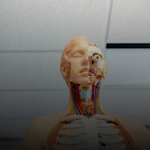
Section 1
Preview this deck
Depressor Labii Inferioris Muscle
Front

| 5 | 0 | ||
| 4 | 0 | ||
| 3 | 0 | ||
| 2 | 0 | ||
| 1 | 0 |
Active users
0
All-time users
0
Favorites
0
Last updated
6 years ago
Date created
Mar 1, 2020
Cards (129)
Section 1
(50 cards)
Depressor Labii Inferioris Muscle
Pulls lower lip down
Risorius Muscle
Joker muscle Elevates corners of mouth
Stylopharyngeus Muscle
THIRD PHARYNGEAL ARCH (GUT TUBE) Styloid process to pharynx Swallowing
Orbicularis Oculi Muscle
Sphincter Closes eyes
Pulp Cavity
Vascular LCT with nerve endings
Crown of Tooth
What you see
Superior Rectus Muscle
Elevates the eyes (upward movement of the eyes)
Buccinator Muscle
Cheek muscles Blowing
Transverse Muscle of Tongue
Projects tongue out Skinny and taller
Procerus Muscle
Depresses eyebrows and wrinkles nose
Nasalis Muscle
Compresses nostrils
Periodontium
Like periosteum Ligament of gomphosis DICT
Zygomaticus Major and Minor
Smiling
Sternocleidomastoid Muscle
LATERAL MESODERM OF THE HEAD Three heads: sternum, clavicle, mastoid Flexes head Contract one: look down to opposite side
Levator Labii Superioris
Elevates upper lip (sneering)
Hyloglossus
depresses and retracts tongue
Neck of Tooth
Above the jaw, below the gum
Digastricus Muscle Posterior Belly
SECOND PHARYNGEAL ARCH (HYOID) Elevates hyoid bone during swallowing
Dentine
Surrounds LCT on all sides Modified bone, mostly hydroxyapatite
Why can't we abduct both eyes?
The brain processes what is in the focal point of vision and cannot split into two fields
Inferior Longitudinal Muscle
Curls tongue under Shortens bottom, elongates top
Pharyngeal and Palatal Muscles
FOURTH PHARYNGEAL ARCH Swallowing Pharynx
Inferior Oblique Muscle
Elevation and Abducts (up and out)
Masseter Muscle
FIRST PHARYNGEAL ARCH (JAW BONES) From zygomatic arch Elevates mandible (biting)
Genioglossus
depresses and protrudes tongue
Laryngeal Muscles
SIXTH PHARYNGEAL ARCH (FIRST OCCIPITAL SOMITE) Move cartilages for speech
Medial Rectus Muscle
Adduction Eyes toward the midline
First Arch Muscles
Temporalis Muscle Masseter Muscle Medial Pterygoid Muscle Lateral Pterygoid Muscle Digastricus Muscle Anterior JAW MUSCLES HAVE BEST MECHANICAL ADVANTAGE IN THE WHOLE BODY
Lateral Pterygoid Muscle
FIRST PHARYNGEAL ARCH (JAW BONES) Attaches mandible Dislocates jaw, protraction Aids in side to side movement
Cement of Tooth
Covers outer surface of root More collagen, 50% hydroxyapatite 40% collagen
Incisors
Flat in the front Cutting
Levator Labii Superioris Alaeque Nasi Muscle
Elevates upper lip and upper side of nose
Superior Oblique Muscle
Depression and Abduction (down and out) Pulley muscle
Occipitofrontalis (frontal belly)
Raises eyebrows
Vertical Muscle of Tongue
Pushes tongue towards the center Elongates tongue
Superior Longitudinal Muscle
Curls tongue up and back Shortens top, lengthens bottom
Temporalis Muscle
FIRST PHARYNGEAL ARCH (JAW BONES) Elevates mandible (closes jaw)
Root of Tooth
Projection into bone
Orbicularis Oris Muscle
Kissing muscle
Mentalis Muscle
Wrinkles chin in concern
Medial Pterygoid Muscle
FIRST PHARYNGEAL ARCH (JAW BONES) Part of sphenoid Runs diagonal Elevates mandible (closes jaw) Contracting one is side to side movement
Platysma Muscle
Lowers mandible Tightens skin Attaches to pectoralis major fascia
Inferior Rectus Muscle
Depresses the eyes (downward movement of the eyes)
Styloglossus
retracts and elevates tongue
Digastricus Muscle Anterior Belly
FIRST PHARYNGEAL ARCH (JAW BONES) Elevates hyoid bone during swallowing
Trapezius
LATERAL MESODERM OF HEAD Elevates head Elevates scapula
Canines
Tall and pointy Grasp and pierce to tear food
Lateral Rectus Muscle
Abducts the eye
Depressor Anguli Oris
Frowning Muscle
Enamel
Surrounds dentin on top Hardest material in vertebrates Doesn't replenish
Section 2
(50 cards)
Cornea
DICT Lets light in and bends it Covers iris
Posterior Chamber
Behind iris and in front of lens
Sphincter Pupillae Muscle
Narrows pupil opening In sun
Optic Disk
Blind spot
Ciliary Muscle
Smooth muscle that alters the shape of the lens for near or far vision
Fovea Centralis
Dimple that increases surface area CONES
Pupil
Black spot of the eye Opening in layer, brings light to lens
Internal Layer of Eye
Retina and optic nerve Allows for light detection
Internal Ear
Fluid filled space with tubes
Sclera
FIBROUS TUNIC White part of the eye Eye muscles pull this
Structure of Temporal Bone
Pyramidal, meets in the middle
Round Window
Secondary tympanic membrane Lower window
Choroid Coat
Vascular LCT Brings blood to eyeball
Iris
Anterior part of middle layer Colored Vascular tissue
Pigmented Epithelium
Cells in the iris Smooth muscle underneath
Parotid Gland
Biggest gland next to the ear
Dilator Pupillae Muscle
Bigger opening In dark
Molars
Back teeth that grind food
Taste Buds
Sensory organs in the mouth Sweet Salty Sour Bitter Umami
Middle Ear
Air-filled space with muscles and bone
Pharyngeal Tonsils (adenoids)
Located in the nasopharynx Immune system and body defense
Vascular Layer of Eye
Vascular LCT Only on eyeball Middle layer
Jugular Floor
Floor of tympanic cavity
Premolars
Cubes, lots of surface area Peaks and valleys Crush food
Vitreous Body
Posterior to lens Gel that maintains shape
Cones
Colors, light
Lens
Focuses light onto retina Transparent behind iris
Filiform Papilla
Most numerous papilla Increase surface area to grasp and manipulate
Conjuctiva
Stratified squamous Continuous with epidermis
Rods
Black, white, grey Low light
Zonular fibers of lens
Thin collagen projections/fibers around lens Change the shape of lens
Fungiform Papilla
Second most numerous Most prominent around edges Allows more area for taste receptors
Lingual Tonsils
Located at the base of the tongue
Aqueous Humor
Fluid in the eye Between the cornea and the lens
Palatine Tonsils
Lateral sides of the tongue (C)
Macula
The central part of the retina containing the fovea
Retina
Holds rods and cones
External Acoustic Meatus
Channel sound waves move through
Ciliary Body
Connects choroid to iris Goes behind lens like a trampoline
Oval Window
Upper window
Dental Formula
2/2 1/1 2/2 3/3 ONE SIDE
Auricle
Side of the head
Fibrous Layer of Eye
Sclera and cornea DICT Covers all eyeball and goes back to brain
Tympanic Membrane
ear drum, separates outer and middle ear Needs equal air on both sides
Anterior Chamber
Between cornea and iris
External Ear
Everything outside Channel to ear
Tegmental Wall
Roof of tympanic cavity
Vallate Papilla
Only 8-12 in mouth Increase surface area for LOTS of taste receptors
Submandibular Gland
Gleak Holds tongue down
Sublingual Gland
Saliva Softens foor for digestion
Section 3
(29 cards)