
Section 1
Preview this deck
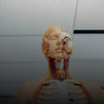
lacrimal none
Front

| 5 | 0 | ||
| 4 | 0 | ||
| 3 | 0 | ||
| 2 | 0 | ||
| 1 | 0 |
Active users
0
All-time users
0
Favorites
0
Last updated
6 years ago
Date created
Mar 1, 2020
Cards (82)
Section 1
(50 cards)
lacrimal none
form medial eye orbit wall. is just beneath the tear gland and duct
vertebral foreman
the opening in each vertebra where the spinal cord passes through
Coronal suture
Suture that joins the parietal and frontal bones
calcaneus
the heel bone; is just inferior to talus
spinous process
the "point" sticking out from the posterior part of a vertebra. these make up the "ridge" of you spine
medial longitudinal arch
the high arch of the foot
Frontal bone
The most anterior of the 3 most superior bones of the body. Forms the forehead
Mandible
lower jaw
Hyiod bone
is the only bone in the body to not be attached to another bone. behind the larynx
ethmoid bone
bone that is most deeply hidden bone of the skull. It forms most of the bony area between the nasal cavity and the eye orbit (socket). Contains some sinuses
Palatine bone
these form anterior hard plate; are part of maxilla; there are 2
zygomatic bone
these form the cheek bone, plus part of eye socket
Sacroliac joint
joint where the sacrum and the ilium articulate
Foramen ovale
opening close to the base of the skull; allows passage of a nerve
External occipital protuberance
The "knot" at the very back of the skull. The point of much muscle attachment
anterior longitudinal ligament, posterior longitudinal ligament
these tough, flexible ligaments run both anterior and posterior along with the full length of the spine; the attach to each vertebrae
talus
foot bone that articulates with the tibia and fibula
medial condyle, lateral condyle
these round, smooth knobs articulate with the tibia. medial is toward the inside
Manibular condyle
important condyle on posterior area of mandible that articulates with the zygomatic process
body
the weight-bearing body of a vertebra; this part rests against intervertebral disc
acetabulum
the area of the hip bone that articulates with the femur, or upper leg bone
occipital condyle
these condyles articulate with the atlas (the 1st vertebra)
vertebral column
the backbone or spine
Supraorbital foramen
a tiny opening above the eye orbit that allows artery and nerve passage
thorasic region
12 vertebrae inferior to the cervical vertebrae
External acoustic meatus
the ear canal; leads to the ear drum
greater trochanter, lesser trochanter
these irregular knobs are areas of muscle attachment, greater is lateral to the head and lesser is inferior to the head
mental foramen
allows blood vessels and nerves to pass from inside jaw to the outside (to lower lip skin and muscle)
Lambdoidal suture
Suture that attaches pariental and occipital bones
Squamous suture
suture that joins the temporal and parietal bone
Mastoid process
the process just posterior to the ear; is the attachment site for several neck muscles
Occipital bone
the most posterior bone of the skull; it also forms most of the base of the skull
Coxal
the two bones that form the hips, or pelvic girdle
shaft
area of rib leading anteriorly to breastbone
styloid process
smaller process medial to the mastoid; attachment site for hyoid bone and several neck muscles
neck
the section of the rib just lateral to head. "curve" in the rib
cervical region
vertebrae numbers 1-7. Vertebra C1 is called "atlas"
Sphenoid bone
a key bone of the cranium; holds all other bones together along the base of the skull; is situated just posterior and slightly superior to the teeth
head
this articulates with the acetabulum of the pelvis
transverse process
the posterior parts of a vertebra; these 2 areas form two arches on the lateral sides of the vertebra, and along with the body, they encircle the spinal cord
medial malleolus
forms the medial "bulge" of the ankle
nasal bone
is the bridge of the nose
zygomatic process
process that extends anteriorly to form the zygomatic arch, which forms the cheekbone
head (rib)
The section of the rib that articulates with a vertebra
Foramen magum
large opening allowing brain stem to pass through to the rest of the brain
Parietal
The most posterior of the 3 most superior bones in the body. They form almost the whole top of the skull
vertabrae
there are 33 of these; together they make up the backbone or spine
lumbar region
last 5
Temporal bone
these bones (are 2) form the area the ear is attached to
lareral malleolous
forms the outside bump on ankle
Section 2
(32 cards)